[English] 日本語
Yorodumi
- PDB-3ric: Crystal Structure of D48V||A47D mutant of Human Glycolipid Transf... -

+
Open data


ID or keywords:

Loading...

-
Basic information

Entry
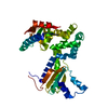
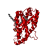
Database: PDB / ID: 3ric
Title
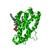
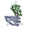
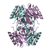
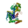
Crystal Structure of D48V||A47D mutant of Human Glycolipid Transfer Protein complexed with 3-O-sulfo-galactosylceramide containing nervonoyl acyl chain (24:1)
ComponentsGlycolipid transfer protein
KeywordsLIPID TRANSPORT / GLTP fold
Function / homology
Function and homology information


glycolipid transfer activity / lipid transfer activity / ceramide transport / ceramide 1-phosphate binding / ceramide 1-phosphate transfer activity / ER to Golgi ceramide transport / Glycosphingolipid transport / glycolipid binding / intermembrane lipid transfer / response to immobilization stress ...glycolipid transfer activity / lipid transfer activity / ceramide transport / ceramide 1-phosphate binding / ceramide 1-phosphate transfer activity / ER to Golgi ceramide transport / Glycosphingolipid transport / glycolipid binding / intermembrane lipid transfer / response to immobilization stress / lipid binding / identical protein binding / cytosol
Similarity search - Function
Glycolipid transfer protein domain / Glycolipid transfer protein superfamily / Glycolipid transfer protein (GLTP) / Glycolipid transfer protein, GLTP / Glycolipid transfer protein / Orthogonal Bundle / Mainly Alpha
Similarity search - Domain/homology
Chem-CIS / Glycolipid transfer protein
Similarity search - Component
Biological speciesHomo sapiens (human)
MethodX-RAY DIFFRACTION / MOLECULAR REPLACEMENT / molecular replacement / Resolution: 2.1 Å
AuthorsSamygina, V. / Ochoa-Lizarralde, B. / Patel, D.J. / Brown, R.E. / Malinina, L.
CitationJournal: Structure / Year: 2011
Title: Enhanced selectivity for sulfatide by engineered human glycolipid transfer protein.
Authors: Samygina, V.R. / Popov, A.N. / Cabo-Bilbao, A. / Ochoa-Lizarralde, B. / Goni-de-Cerio, F. / Zhai, X. / Molotkovsky, J.G. / Patel, D.J. / Brown, R.E. / Malinina, L.
History
DepositionApr 13, 2011Deposition site: RCSB / Processing site: RCSB
Revision 1.0Feb 8, 2012Provider: repository / Type: Initial release
Revision 1.1Jun 20, 2012Group: Data collection
Revision 1.2Nov 8, 2017Group: Refinement description / Category: software / Item: _software.name
Revision 1.3Feb 28, 2024Group: Data collection / Database references / Derived calculations
Category: chem_comp_atom / chem_comp_bond ...chem_comp_atom / chem_comp_bond / database_2 / struct_ref_seq_dif / struct_site
Item: _database_2.pdbx_DOI / _database_2.pdbx_database_accession ..._database_2.pdbx_DOI / _database_2.pdbx_database_accession / _struct_ref_seq_dif.details / _struct_site.pdbx_auth_asym_id / _struct_site.pdbx_auth_comp_id / _struct_site.pdbx_auth_seq_id

-
Structure visualization

Structure viewerMolecule:
MolmilJmol/JSmol

Downloads & links

-
Assembly

Deposited unit
A: Glycolipid transfer protein
hetero molecules


Theoretical massNumber of molelcules
Total (without water)24,7962
Polymers23,9061
Non-polymers8901
Water1,13563
1
A: Glycolipid transfer protein
hetero molecules

A: Glycolipid transfer protein
hetero molecules


Theoretical massNumber of molelcules
Total (without water)49,5924
Polymers47,8122
Non-polymers1,7812
Water362
TypeNameSymmetry operationNumber
identity operation1_555x,y,z1
crystal symmetry operation2_454-x-1,y,-z-11
Buried area5580 Å2
ΔGint-36 kcal/mol
Surface area19390 Å2
MethodPISA
Unit cell
Length a, b, c (Å)76.871, 48.128, 69.086
Angle α, β, γ (deg.)90.00, 123.82, 90.00
Int Tables number5
Space group name H-MC121

-
Components

#1: Protein Glycolipid transfer protein / GLTP


Mass: 23905.830 Da / Num. of mol.: 1 / Fragment: Human Glycolipid Transfer Protein / Mutation: D48V, A47D
Source method: isolated from a genetically manipulated source
Source: (gene. exp.) Homo sapiens (human) / Gene: GLTP / Production host: Escherichia coli (E. coli) / References: UniProt: Q9NZD2, DNA-directed DNA polymerase
#2: Chemical ChemComp-CIS / (15Z)-N-((1S,2R,3E)-2-HYDROXY-1-{[(3-O-SULFO-BETA-D-GALACTOPYRANOSYL)OXY]METHYL}HEPTADEC-3-ENYL)TETRACOS-15-ENAMIDE / (2S,3R,4E)-N-NERVONIC-1-[BETA-D-(3-SULFATE)-GALACTOPYRANOSYL]-2-AMINO-OCTADECENE-3-OL / CIS-TETRACOSENOYL SULFATIDE


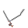
Mass: 890.301 Da / Num. of mol.: 1 / Source method: obtained synthetically / Formula: C48H91NO11S
#3: Water ChemComp-HOH / water


Mass: 18.015 Da / Num. of mol.: 63 / Source method: isolated from a natural source / Formula: H2O

-
Experimental details

-
Experiment

ExperimentMethod: X-RAY DIFFRACTION / Number of used crystals: 1

-
Sample preparation

CrystalDensity Matthews: 2.22 Å3/Da / Density % sol: 44.61 %
Crystal growTemperature: 273 K / pH: 5.1
Details: 15-20% PEG 3350, pH 5.1, VAPOR DIFFUSION, HANGING DROP, temperature 273K

-
Data collection

DiffractionMean temperature: 100 K
Diffraction sourceSource: ROTATING ANODE / Type: BRUKER AXS MICROSTAR / Wavelength: 1.54
DetectorType: MAR scanner 345 mm plate / Detector: IMAGE PLATE / Date: Mar 22, 2011
RadiationProtocol: SINGLE WAVELENGTH / Monochromatic (M) / Laue (L): M / Scattering type: x-ray
Radiation wavelengthWavelength: 1.54 Å / Relative weight: 1
ReflectionResolution: 2.1→20 Å / Num. obs: 11907 / % possible obs: 96 % / Redundancy: 4.6 % / Rmerge(I) obs: 0.05 / Net I/σ(I): 14.2
Reflection shellResolution: 2.1→2.17 Å / Redundancy: 4.6 % / Rmerge(I) obs: 0.55 / % possible all: 93.7

-
Phasing

PhasingMethod: molecular replacement

-
Processing

Software
NameVersionClassificationNB
SCALEPACKdata scaling
MOLREPphasing
REFMAC5.2.0019refinement
PDB_EXTRACT3.1data extraction
DENZOdata reduction
RefinementMethod to determine structure: MOLECULAR REPLACEMENT / Resolution: 2.1→14.98 Å / Cor.coef. Fo:Fc: 0.969 / Cor.coef. Fo:Fc free: 0.958 / Occupancy max: 1 / Occupancy min: 0.5 / SU B: 13.717 / SU ML: 0.16 / Cross valid method: THROUGHOUT / ESU R: 0.262 / ESU R Free: 0.189 / Stereochemistry target values: MAXIMUM LIKELIHOOD / Details: HYDROGENS HAVE BEEN ADDED IN THE RIDING POSITIONS
RfactorNum. reflection% reflectionSelection details
Rfree0.21881 570 4.8 %RANDOM
Rwork0.18191 ---
obs0.18378 11300 96.08 %-
Solvent computationIon probe radii: 0.8 Å / Shrinkage radii: 0.8 Å / VDW probe radii: 1.2 Å / Solvent model: MASK
Displacement parametersBiso mean: 51.935 Å2
Baniso -1Baniso -2Baniso -3
1--0.47 Å20 Å2-0.14 Å2
2--0.23 Å20 Å2
3---0.09 Å2
Refine analyze
FreeObs
Luzzati coordinate error0.195 Å0.288 Å
Refinement stepCycle: LAST / Resolution: 2.1→14.98 Å
ProteinNucleic acidLigandSolventTotal
Num. atoms1670 0 61 63 1794
Refine LS restraints
Refine-IDTypeDev idealDev ideal targetNumber
X-RAY DIFFRACTIONr_bond_refined_d0.0170.0221781
X-RAY DIFFRACTIONr_bond_other_d0.0010.021257
X-RAY DIFFRACTIONr_angle_refined_deg1.6582.0042403
X-RAY DIFFRACTIONr_angle_other_deg0.96233090
X-RAY DIFFRACTIONr_dihedral_angle_1_deg6.3225206
X-RAY DIFFRACTIONr_dihedral_angle_2_deg32.38924.60576
X-RAY DIFFRACTIONr_dihedral_angle_3_deg16.18215313
X-RAY DIFFRACTIONr_dihedral_angle_4_deg22.258157
X-RAY DIFFRACTIONr_chiral_restr0.0950.2269
X-RAY DIFFRACTIONr_gen_planes_refined0.0060.021877
X-RAY DIFFRACTIONr_gen_planes_other0.0010.02340
X-RAY DIFFRACTIONr_nbd_refined0.2150.2445
X-RAY DIFFRACTIONr_nbd_other0.1870.21238
X-RAY DIFFRACTIONr_nbtor_refined0.1860.2865
X-RAY DIFFRACTIONr_nbtor_other0.0890.2860
X-RAY DIFFRACTIONr_xyhbond_nbd_refined0.1510.256
X-RAY DIFFRACTIONr_xyhbond_nbd_other
X-RAY DIFFRACTIONr_metal_ion_refined
X-RAY DIFFRACTIONr_metal_ion_other
X-RAY DIFFRACTIONr_symmetry_vdw_refined0.1760.223
X-RAY DIFFRACTIONr_symmetry_vdw_other0.2420.241
X-RAY DIFFRACTIONr_symmetry_hbond_refined0.2130.26
X-RAY DIFFRACTIONr_symmetry_hbond_other
X-RAY DIFFRACTIONr_symmetry_metal_ion_refined
X-RAY DIFFRACTIONr_symmetry_metal_ion_other
X-RAY DIFFRACTIONr_mcbond_it0.9671.51352
X-RAY DIFFRACTIONr_mcbond_other0.2141.5411
X-RAY DIFFRACTIONr_mcangle_it1.18621685
X-RAY DIFFRACTIONr_scbond_it2.0313875
X-RAY DIFFRACTIONr_scangle_it2.8494.5718
X-RAY DIFFRACTIONr_rigid_bond_restr
X-RAY DIFFRACTIONr_sphericity_free
X-RAY DIFFRACTIONr_sphericity_bonded
LS refinement shellResolution: 2.1→2.153 Å / Total num. of bins used: 20
RfactorNum. reflection% reflection
Rfree0.341 41 -
Rwork0.257 812 -
obs--93.84 %
Refinement TLS params.Method: refined / Origin x: -10.7349 Å / Origin y: -0.8487 Å / Origin z: -14.9749 Å
111213212223313233
T-0.0834 Å20.0077 Å20.0053 Å2--0.1527 Å20.0672 Å2---0.1791 Å2
L3.6979 °2-1.1342 °20.9295 °2-2.8786 °20.4038 °2--1.6891 °2
S0.1456 Å °-0.2158 Å °-0.276 Å °-0.2829 Å °0.0401 Å °-0.09 Å °-0.0146 Å °0.0287 Å °-0.1857 Å °
Refinement TLS group
IDRefine-IDRefine TLS-IDAuth asym-IDAuth seq-ID
1X-RAY DIFFRACTION1A401 - 463
2X-RAY DIFFRACTION1A301
3X-RAY DIFFRACTION1A3 - 209

+
About Yorodumi

-
News

-
Feb 9, 2022. New format data for meta-information of EMDB entries

New format data for meta-information of EMDB entries

  • Version 3 of the EMDB header file is now the official format.
  • The previous official version 1.9 will be removed from the archive.

Related info.:EMDB header

External links:wwPDB to switch to version 3 of the EMDB data model

-
Aug 12, 2020. Covid-19 info

Covid-19 info

URL: https://pdbjlvh1.pdbj.org/emnavi/covid19.php

New page: Covid-19 featured information page in EM Navigator.

Related info.:Covid-19 info / Mar 5, 2020. Novel coronavirus structure data

+
Mar 5, 2020. Novel coronavirus structure data

Novel coronavirus structure data

Related info.:Yorodumi Speices / Aug 12, 2020. Covid-19 info

External links:COVID-19 featured content - PDBj / Molecule of the Month (242):Coronavirus Proteases

+
Jan 31, 2019. EMDB accession codes are about to change! (news from PDBe EMDB page)

EMDB accession codes are about to change! (news from PDBe EMDB page)

  • The allocation of 4 digits for EMDB accession codes will soon come to an end. Whilst these codes will remain in use, new EMDB accession codes will include an additional digit and will expand incrementally as the available range of codes is exhausted. The current 4-digit format prefixed with “EMD-” (i.e. EMD-XXXX) will advance to a 5-digit format (i.e. EMD-XXXXX), and so on. It is currently estimated that the 4-digit codes will be depleted around Spring 2019, at which point the 5-digit format will come into force.
  • The EM Navigator/Yorodumi systems omit the EMD- prefix.

Related info.:Q: What is EMD? / ID/Accession-code notation in Yorodumi/EM Navigator

External links:EMDB Accession Codes are Changing Soon! / Contact to PDBj

+
Jul 12, 2017. Major update of PDB

Major update of PDB

  • wwPDB released updated PDB data conforming to the new PDBx/mmCIF dictionary.
  • This is a major update changing the version number from 4 to 5, and with Remediation, in which all the entries are updated.
  • In this update, many items about electron microscopy experimental information are reorganized (e.g. em_software).
  • Now, EM Navigator and Yorodumi are based on the updated data.

External links:wwPDB Remediation / Enriched Model Files Conforming to OneDep Data Standards Now Available in the PDB FTP Archive

-
Yorodumi

Thousand views of thousand structures

  • Yorodumi is a browser for structure data from EMDB, PDB, SASBDB, etc.
  • This page is also the successor to EM Navigator detail page, and also detail information page/front-end page for Omokage search.
  • The word "yorodu" (or yorozu) is an old Japanese word meaning "ten thousand". "mi" (miru) is to see.

Related info.:EMDB / PDB / SASBDB / Comparison of 3 databanks / Yorodumi Search / Aug 31, 2016. New EM Navigator & Yorodumi / Yorodumi Papers / Jmol/JSmol / Function and homology information / Changes in new EM Navigator and Yorodumi

Read more