 Movie
Movie Controller

[English] 日本語
 Yorodumi
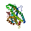
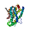
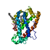
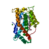
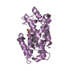
- PDB-2eum: Crystal structure of human Glycolipid Transfer Protein complexed ... -
+ Open data

- Basic information
| Entry | Database: PDB / ID: 2eum | |||||||||
|---|---|---|---|---|---|---|---|---|---|---|
| Title | Crystal structure of human Glycolipid Transfer Protein complexed with 8:0 Lactosylceramide | |||||||||
 Components Components | Glycolipid transfer protein | |||||||||
 Keywords Keywords | LIPID TRANSPORT / Protein-glycolipid complex | |||||||||
| Function / homology |  Function and homology information Function and homology informationglycolipid transfer activity / ceramide 1-phosphate transfer activity / ceramide transport / ceramide 1-phosphate binding / glycolipid binding / lipid transfer activity / Glycosphingolipid transport / intermembrane lipid transfer / response to immobilization stress / lipid binding ...glycolipid transfer activity / ceramide 1-phosphate transfer activity / ceramide transport / ceramide 1-phosphate binding / glycolipid binding / lipid transfer activity / Glycosphingolipid transport / intermembrane lipid transfer / response to immobilization stress / lipid binding / identical protein binding / cytosol Similarity search - Function | |||||||||
| Biological species |  Homo sapiens (human) Homo sapiens (human) | |||||||||
| Method |  X-RAY DIFFRACTION / MOLECULAR REPLACEMENT / Resolution: 2.3 Å X-RAY DIFFRACTION / MOLECULAR REPLACEMENT / Resolution: 2.3 Å | |||||||||
 Authors Authors | Malinina, L. / Malakhova, M.L. / Kanack, A.T. / Abagyan, R. / Brown, R.E. / Patel, D.J. | |||||||||
 Citation Citation | Journal: Plos Biol. / Year: 2006 Title: The liganding of glycolipid transfer protein is controlled by glycolipid acyl structure. Authors: Malinina, L. / Malakhova, M.L. / Kanack, A.T. / Lu, M. / Abagyan, R. / Brown, R.E. / Patel, D.J. | |||||||||
| History |
|
- Structure visualization
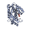
| Structure viewer | Molecule: MolmilJmol/JSmol |
|---|
- Downloads & links
-Download
| PDBx/mmCIF format | 2eum.cif.gz | 62.9 KB | Display | PDBx/mmCIF format |
|---|---|---|---|---|
| PDB format | pdb2eum.ent.gz | 43.1 KB | Display | PDB format |
| PDBx/mmJSON format | 2eum.json.gz | Tree view | PDBx/mmJSON format | |
| Others |  Other downloads Other downloads |
-Validation report
| Arichive directory | https://data.pdbj.org/pub/pdb/validation_reports/eu/2eumftp://data.pdbj.org/pub/pdb/validation_reports/eu/2eum | HTTPS FTP |
|---|
-Related structure data
| Related structure data |  2eukSC  2evdC 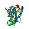 2evlC 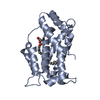 2evsC  2evtC S: Starting model for refinement C: citing same article ( |
|---|---|
| Similar structure data |


-Links
 PDBj
PDBj


- Assembly
| Deposited unit | 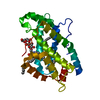
| ||||||||
|---|---|---|---|---|---|---|---|---|---|
| 1 |
| ||||||||
| Unit cell |
| ||||||||
| Components on special symmetry positions |
|
-Components
-Protein / Sugars , 2 types, 2 molecules A
| #1: Protein | Mass: 23877.777 Da / Num. of mol.: 1 Source method: isolated from a genetically manipulated source Source: (gene. exp.) Homo sapiens (human) / Plasmid: PET30 XA-LIC / Production host:  |
|---|---|
| #2: Polysaccharide | beta-D-galactopyranose-(1-4)-beta-D-glucopyranose / beta-lactose  Source method: isolated from a genetically manipulated source Details: oligosaccharide / References: beta-lactose |
-Non-polymers , 5 types, 157 molecules 


| #3: Chemical | ChemComp-SPH /  Mass: 299.492 Da / Num. of mol.: 1 / Source method: obtained synthetically / Formula: C18H37NO2 Mass: 299.492 Da / Num. of mol.: 1 / Source method: obtained synthetically / Formula: C18H37NO2 |
|---|---|
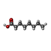
| #4: Chemical | ChemComp-OCA /  Mass: 144.211 Da / Num. of mol.: 1 / Source method: obtained synthetically / Formula: C8H16O2 Mass: 144.211 Da / Num. of mol.: 1 / Source method: obtained synthetically / Formula: C8H16O2 |
| #5: Chemical | ChemComp-OCT /  Mass: 114.229 Da / Num. of mol.: 1 / Source method: obtained synthetically / Formula: C8H18 Mass: 114.229 Da / Num. of mol.: 1 / Source method: obtained synthetically / Formula: C8H18 |
| #6: Chemical | ChemComp-D10 /  Mass: 142.282 Da / Num. of mol.: 1 / Source method: obtained synthetically / Formula: C10H22 Mass: 142.282 Da / Num. of mol.: 1 / Source method: obtained synthetically / Formula: C10H22 |
| #7: Water | ChemComp-HOH / Mass: 18.015 Da / Num. of mol.: 153 / Source method: isolated from a natural source / Formula: H2O |
-Experimental details
-Experiment
| Experiment | Method: X-RAY DIFFRACTION / Number of used crystals: 1 |
|---|
- Sample preparation
| Crystal | Density Matthews: 2.25 Å3/Da / Density % sol: 45.39 % |
|---|---|
| Crystal grow | Temperature: 293 K / Method: vapor diffusion / pH: 4.5 Details: 15-20% PEG 3350 or 8000, 50 mM possium phosphate, pH 4.5, VAPOR DIFFUSION, temperature 293K |
-Data collection
| Diffraction | Mean temperature: 100 K |
|---|---|
| Diffraction source | Source: ROTATING ANODE / Type: RIGAKU RUH3R / Wavelength: 1.5418 Å |
| Detector | Type: RIGAKU RAXIS IV / Detector: IMAGE PLATE / Date: Oct 28, 2005 / Details: mirrors |
| Radiation | Protocol: SINGLE WAVELENGTH / Monochromatic (M) / Laue (L): M / Scattering type: x-ray |
| Radiation wavelength | Wavelength: 1.5418 Å / Relative weight: 1 |
| Reflection | Resolution: 2.3→20 Å / Num. all: 9584 / Num. obs: 9562 / % possible obs: 99.9 % / Observed criterion σ(F): 0 / Observed criterion σ(I): 0 / Redundancy: 3.6 % / Rmerge(I) obs: 0.095 / Χ2: 1.195 |
| Reflection shell | Resolution: 2.3→2.38 Å / Redundancy: 3.4 % / Rmerge(I) obs: 0.523 / Num. unique all: 946 / Χ2: 1.481 / % possible all: 99.9 |
- Processing
| Software |
| ||||||||||||||||||||||||||||||||||||||||||||||||||||||||||||||||||||||||||||||||||||||||||
|---|---|---|---|---|---|---|---|---|---|---|---|---|---|---|---|---|---|---|---|---|---|---|---|---|---|---|---|---|---|---|---|---|---|---|---|---|---|---|---|---|---|---|---|---|---|---|---|---|---|---|---|---|---|---|---|---|---|---|---|---|---|---|---|---|---|---|---|---|---|---|---|---|---|---|---|---|---|---|---|---|---|---|---|---|---|---|---|---|---|---|---|
| Refinement | Method to determine structure: MOLECULAR REPLACEMENT Starting model: PDB ENTRY 2EUK Resolution: 2.3→20 Å / Cor.coef. Fo:Fc: 0.959 / Cor.coef. Fo:Fc free: 0.921 / SU B: 8.634 / SU ML: 0.205 / Cross valid method: THROUGHOUT / σ(F): 0 / ESU R: 0.462 / ESU R Free: 0.266 / Stereochemistry target values: MAXIMUM LIKELIHOOD / Details: HYDROGENS HAVE BEEN ADDED IN THE RIDING POSITIONS
| ||||||||||||||||||||||||||||||||||||||||||||||||||||||||||||||||||||||||||||||||||||||||||
| Solvent computation | Ion probe radii: 0.8 Å / Shrinkage radii: 0.8 Å / VDW probe radii: 1.2 Å / Solvent model: MASK | ||||||||||||||||||||||||||||||||||||||||||||||||||||||||||||||||||||||||||||||||||||||||||
| Displacement parameters | Biso mean: 43.892 Å2
| ||||||||||||||||||||||||||||||||||||||||||||||||||||||||||||||||||||||||||||||||||||||||||
| Refinement step | Cycle: LAST / Resolution: 2.3→20 Å
| ||||||||||||||||||||||||||||||||||||||||||||||||||||||||||||||||||||||||||||||||||||||||||
| Refine LS restraints |
| ||||||||||||||||||||||||||||||||||||||||||||||||||||||||||||||||||||||||||||||||||||||||||
| LS refinement shell | Resolution: 2.3→2.359 Å / Total num. of bins used: 20
|
