 Movie
Movie Controller

[English] 日本語
 Yorodumi
- PDB-5k3e: Crystal Structure of the Fluoroacetate Dehalogenase RPA1163 - Asp... -
+ Open data

- Basic information
| Entry | Database: PDB / ID: 5k3e | |||||||||
|---|---|---|---|---|---|---|---|---|---|---|
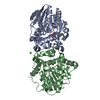
| Title | Crystal Structure of the Fluoroacetate Dehalogenase RPA1163 - Asp110Asn/Glycolate - Cocrystallized | |||||||||
 Components Components | Fluoroacetate dehalogenase | |||||||||
 Keywords Keywords | HYDROLASE / Homodimer / Dehalogenase | |||||||||
| Function / homology |  Function and homology information Function and homology information | |||||||||
| Biological species |  Rhodopseudomonas palustris (phototrophic) Rhodopseudomonas palustris (phototrophic) | |||||||||
| Method |  X-RAY DIFFRACTION / MOLECULAR REPLACEMENT / Resolution: 1.54 Å X-RAY DIFFRACTION / MOLECULAR REPLACEMENT / Resolution: 1.54 Å | |||||||||
 Authors Authors | Mehrabi, P. / Kim, T.H. / Prosser, S.R. / Pai, E.F. | |||||||||
 Citation Citation | Journal: Science / Year: 2017 Title: The role of dimer asymmetry and protomer dynamics in enzyme catalysis. Authors: Kim, T.H. / Mehrabi, P. / Ren, Z. / Sljoka, A. / Ing, C. / Bezginov, A. / Ye, L. / Pomes, R. / Prosser, R.S. / Pai, E.F. | |||||||||
| History |
|
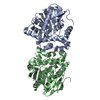
- Structure visualization
| Structure viewer | Molecule: MolmilJmol/JSmol |
|---|
- Downloads & links
-Download
| PDBx/mmCIF format | 5k3e.cif.gz | 145.9 KB | Display | PDBx/mmCIF format |
|---|---|---|---|---|
| PDB format | pdb5k3e.ent.gz | 112 KB | Display | PDB format |
| PDBx/mmJSON format | 5k3e.json.gz | Tree view | PDBx/mmJSON format | |
| Others |  Other downloads Other downloads |
-Validation report
| Arichive directory | https://data.pdbj.org/pub/pdb/validation_reports/k3/5k3eftp://data.pdbj.org/pub/pdb/validation_reports/k3/5k3e | HTTPS FTP |
|---|
-Related structure data
| Related structure data |  5k3aC  5k3bC  5k3cC  5k3dC 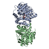 5k3fC 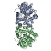 5swnC 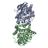 5t4tC 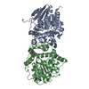 3r3uS C: citing same article ( S: Starting model for refinement |
|---|---|
| Similar structure data |


-Links
 PDBj
PDBj


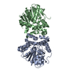
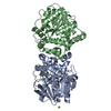
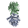
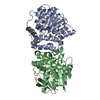
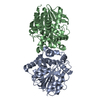
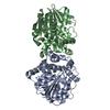
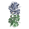
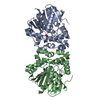
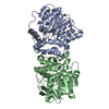
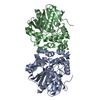
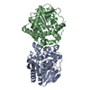
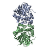
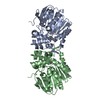
- Assembly
| Deposited unit | 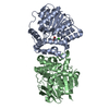
| ||||||||
|---|---|---|---|---|---|---|---|---|---|
| 1 |
| ||||||||
| Unit cell |
|
-Components
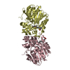
| #1: Protein | Mass: 34072.676 Da / Num. of mol.: 2 / Mutation: D110N Source method: isolated from a genetically manipulated source Source: (gene. exp.) Rhodopseudomonas palustris (strain ATCC BAA-98 / CGA009) (phototrophic)Strain: ATCC BAA-98 / CGA009 / Gene: RPA1163 / Production host: #2: Chemical | ChemComp-GOA / | 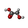  Mass: 76.051 Da / Num. of mol.: 1 / Source method: obtained synthetically / Formula: C2H4O3 Mass: 76.051 Da / Num. of mol.: 1 / Source method: obtained synthetically / Formula: C2H4O3#3: Chemical |   Mass: 35.453 Da / Num. of mol.: 2 / Source method: obtained synthetically / Formula: Cl Mass: 35.453 Da / Num. of mol.: 2 / Source method: obtained synthetically / Formula: Cl#4: Water | ChemComp-HOH / |  Mass: 18.015 Da / Num. of mol.: 648 / Source method: isolated from a natural source / Formula: H2O Mass: 18.015 Da / Num. of mol.: 648 / Source method: isolated from a natural source / Formula: H2O |
|---|
-Experimental details
-Experiment
| Experiment | Method: X-RAY DIFFRACTION / Number of used crystals: 1 |
|---|
- Sample preparation
| Crystal | Density Matthews: 2.06 Å3/Da / Density % sol: 40.27 % |
|---|---|
| Crystal grow | Temperature: 298 K / Method: vapor diffusion, hanging drop / pH: 8.5 Details: PEG 3350 19-22%, 200mM Calcium chloride, 100mM Tris HCL pH 8.5 |
-Data collection
| Diffraction | Mean temperature: 93 K |
|---|---|
| Diffraction source | Source: ROTATING ANODE / Type: RIGAKU FR-D / Wavelength: 1.5418 Å |
| Detector | Type: MAR scanner 345 mm plate / Detector: IMAGE PLATE / Date: Nov 11, 2013 |
| Radiation | Protocol: SINGLE WAVELENGTH / Monochromatic (M) / Laue (L): M / Scattering type: x-ray |
| Radiation wavelength | Wavelength: 1.5418 Å / Relative weight: 1 |
| Reflection | Resolution: 1.54→19.8 Å / Num. obs: 78608 / % possible obs: 99.5 % / Redundancy: 2 % / Rmerge(I) obs: 0.024 / Net I/σ(I): 24 |
| Reflection shell | Resolution: 1.54→1.58 Å / Rmerge(I) obs: 0.17 |
- Processing
| Software |
| |||||||||||||||||||||||||||||||||||||||||||||||||||||||||||||||||||||||||||||||||||||||||||||||||||||||||||||||||||||||||||||||||||||||||||||||||||||||||||||||||||||||||||||||||||||||||||||||||||||||||||
|---|---|---|---|---|---|---|---|---|---|---|---|---|---|---|---|---|---|---|---|---|---|---|---|---|---|---|---|---|---|---|---|---|---|---|---|---|---|---|---|---|---|---|---|---|---|---|---|---|---|---|---|---|---|---|---|---|---|---|---|---|---|---|---|---|---|---|---|---|---|---|---|---|---|---|---|---|---|---|---|---|---|---|---|---|---|---|---|---|---|---|---|---|---|---|---|---|---|---|---|---|---|---|---|---|---|---|---|---|---|---|---|---|---|---|---|---|---|---|---|---|---|---|---|---|---|---|---|---|---|---|---|---|---|---|---|---|---|---|---|---|---|---|---|---|---|---|---|---|---|---|---|---|---|---|---|---|---|---|---|---|---|---|---|---|---|---|---|---|---|---|---|---|---|---|---|---|---|---|---|---|---|---|---|---|---|---|---|---|---|---|---|---|---|---|---|---|---|---|---|---|---|---|---|---|
| Refinement | Method to determine structure: MOLECULAR REPLACEMENT Starting model: 3R3U Resolution: 1.54→19.776 Å / SU ML: 0.16 / Cross valid method: FREE R-VALUE / σ(F): 1.34 / Phase error: 20.59
| |||||||||||||||||||||||||||||||||||||||||||||||||||||||||||||||||||||||||||||||||||||||||||||||||||||||||||||||||||||||||||||||||||||||||||||||||||||||||||||||||||||||||||||||||||||||||||||||||||||||||||
| Solvent computation | Shrinkage radii: 0.9 Å / VDW probe radii: 1.11 Å | |||||||||||||||||||||||||||||||||||||||||||||||||||||||||||||||||||||||||||||||||||||||||||||||||||||||||||||||||||||||||||||||||||||||||||||||||||||||||||||||||||||||||||||||||||||||||||||||||||||||||||
| Displacement parameters | Biso max: 64.89 Å2 / Biso mean: 16.0274 Å2 / Biso min: 4.53 Å2 | |||||||||||||||||||||||||||||||||||||||||||||||||||||||||||||||||||||||||||||||||||||||||||||||||||||||||||||||||||||||||||||||||||||||||||||||||||||||||||||||||||||||||||||||||||||||||||||||||||||||||||
| Refinement step | Cycle: final / Resolution: 1.54→19.776 Å
| |||||||||||||||||||||||||||||||||||||||||||||||||||||||||||||||||||||||||||||||||||||||||||||||||||||||||||||||||||||||||||||||||||||||||||||||||||||||||||||||||||||||||||||||||||||||||||||||||||||||||||
| Refine LS restraints |
| |||||||||||||||||||||||||||||||||||||||||||||||||||||||||||||||||||||||||||||||||||||||||||||||||||||||||||||||||||||||||||||||||||||||||||||||||||||||||||||||||||||||||||||||||||||||||||||||||||||||||||
| LS refinement shell | Refine-ID: X-RAY DIFFRACTION / Total num. of bins used: 28
|
