 Movie
Movie Controller

[English] 日本語
 Yorodumi
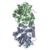
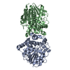
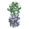
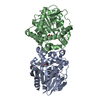
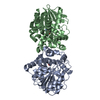
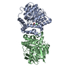
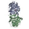
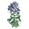
- PDB-7a42: Fluoroacetate Dehalogenase measured by serial synchrotron crystal... -
+ Open data

- Basic information
| Entry | Database: PDB / ID: 7a42 | ||||||
|---|---|---|---|---|---|---|---|
| Title | Fluoroacetate Dehalogenase measured by serial synchrotron crystallography | ||||||
 Components Components | Fluoroacetate dehalogenase | ||||||
 Keywords Keywords | HYDROLASE / dehalogenase / serial synchrotron crystallography | ||||||
| Function / homology | haloacetate dehalogenase / haloacetate dehalogenase activity / Epoxide hydrolase-like / alpha/beta hydrolase fold / Alpha/beta hydrolase fold-1 / Alpha/Beta hydrolase fold / Fluoroacetate dehalogenase Function and homology information Function and homology information | ||||||
| Biological species |  Rhodopseudomonas palustris (phototrophic) Rhodopseudomonas palustris (phototrophic) | ||||||
| Method |  X-RAY DIFFRACTION / SYNCHROTRON / MOLECULAR REPLACEMENT / Resolution: 1.75 Å X-RAY DIFFRACTION / SYNCHROTRON / MOLECULAR REPLACEMENT / Resolution: 1.75 Å | ||||||
 Authors Authors | Mehrabi, P. / Schulz, E.C. / Buecker, R. | ||||||
 Citation Citation | Journal: Sci Adv / Year: 2021 Title: Serial femtosecond and serial synchrotron crystallography can yield data of equivalent quality: A systematic comparison. Authors: Mehrabi, P. / Bucker, R. / Bourenkov, G. / Ginn, H.M. / von Stetten, D. / Muller-Werkmeister, H.M. / Kuo, A. / Morizumi, T. / Eger, B.T. / Ou, W.L. / Oghbaey, S. / Sarracini, A. / Besaw, J.E. ...Authors: Mehrabi, P. / Bucker, R. / Bourenkov, G. / Ginn, H.M. / von Stetten, D. / Muller-Werkmeister, H.M. / Kuo, A. / Morizumi, T. / Eger, B.T. / Ou, W.L. / Oghbaey, S. / Sarracini, A. / Besaw, J.E. / Pare-Labrosse, O. / Meier, S. / Schikora, H. / Tellkamp, F. / Marx, A. / Sherrell, D.A. / Axford, D. / Owen, R.L. / Ernst, O.P. / Pai, E.F. / Schulz, E.C. / Miller, R.J.D. | ||||||
| History |
|
- Structure visualization
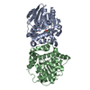
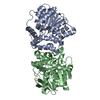
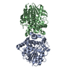
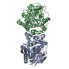
| Structure viewer | Molecule: MolmilJmol/JSmol |
|---|
- Downloads & links
-Download
| PDBx/mmCIF format | 7a42.cif.gz | 167.3 KB | Display | PDBx/mmCIF format |
|---|---|---|---|---|
| PDB format | pdb7a42.ent.gz | 106.5 KB | Display | PDB format |
| PDBx/mmJSON format | 7a42.json.gz | Tree view | PDBx/mmJSON format | |
| Others |  Other downloads Other downloads |
-Validation report
| Arichive directory | https://data.pdbj.org/pub/pdb/validation_reports/a4/7a42ftp://data.pdbj.org/pub/pdb/validation_reports/a4/7a42 | HTTPS FTP |
|---|
-Related structure data
| Related structure data | 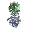 7a43C 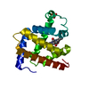 7a44C 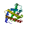 7a45C 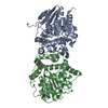 3r3uS S: Starting model for refinement C: citing same article ( |
|---|---|
| Similar structure data |


-Links
 PDBj
PDBj


- Assembly
| Deposited unit | 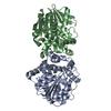
| ||||||||||||
|---|---|---|---|---|---|---|---|---|---|---|---|---|---|
| 1 |
| ||||||||||||
| Unit cell |
|
-Components
| #1: Protein | Mass: 34073.660 Da / Num. of mol.: 2 Source method: isolated from a genetically manipulated source Source: (gene. exp.) Rhodopseudomonas palustris (phototrophic)Gene: RPA1163 / Production host: #2: Chemical |   Mass: 35.453 Da / Num. of mol.: 2 / Source method: obtained synthetically / Formula: Cl / Feature type: SUBJECT OF INVESTIGATION Mass: 35.453 Da / Num. of mol.: 2 / Source method: obtained synthetically / Formula: Cl / Feature type: SUBJECT OF INVESTIGATION#3: Water | ChemComp-HOH / |  Mass: 18.015 Da / Num. of mol.: 300 / Source method: isolated from a natural source / Formula: H2O Mass: 18.015 Da / Num. of mol.: 300 / Source method: isolated from a natural source / Formula: H2OHas ligand of interest | Y | |
|---|
-Experimental details
-Experiment
| Experiment | Method: X-RAY DIFFRACTION / Number of used crystals: 1 |
|---|
- Sample preparation
| Crystal | Density Matthews: 2.03 Å3/Da / Density % sol: 39.3 % |
|---|---|
| Crystal grow | Temperature: 293 K / Method: batch mode Details: 18-20 % (w/v)) PEG3350, 200 mM CaCl2, and 100 mM Tris-HCl pH 8.5 |
-Data collection
| Diffraction | Mean temperature: 293 K / Serial crystal experiment: Y |
|---|---|
| Diffraction source | Source: SYNCHROTRON / Site: PETRA III, EMBL c/o DESY  / Beamline: P14 (MX2) / Wavelength: 0.9778 Å / Beamline: P14 (MX2) / Wavelength: 0.9778 Å |
| Detector | Type: DECTRIS PILATUS 6M / Detector: PIXEL / Date: May 27, 2016 |
| Radiation | Protocol: SINGLE WAVELENGTH / Monochromatic (M) / Laue (L): M / Scattering type: x-ray |
| Radiation wavelength | Wavelength: 0.9778 Å / Relative weight: 1 |
| Reflection | Resolution: 1.75→57.39 Å / Num. obs: 54827 / % possible obs: 100 % / Redundancy: 139.52 % / Biso Wilson estimate: 18.85 Å2 / CC1/2: 0.9534 / Net I/σ(I): 4.18 |
| Reflection shell | Resolution: 1.75→1.813 Å / Num. unique obs: 5456 / CC1/2: 0.5252 |
| Serial crystallography sample delivery | Method: fixed target |
- Processing
| Software |
| ||||||||||||||||||||||||||||||||||||||||||||||||||||||||||||||||||||||||||||||||||||||||||||||||||
|---|---|---|---|---|---|---|---|---|---|---|---|---|---|---|---|---|---|---|---|---|---|---|---|---|---|---|---|---|---|---|---|---|---|---|---|---|---|---|---|---|---|---|---|---|---|---|---|---|---|---|---|---|---|---|---|---|---|---|---|---|---|---|---|---|---|---|---|---|---|---|---|---|---|---|---|---|---|---|---|---|---|---|---|---|---|---|---|---|---|---|---|---|---|---|---|---|---|---|---|
| Refinement | Method to determine structure: MOLECULAR REPLACEMENT Starting model: 3r3u Resolution: 1.75→33.08 Å / SU ML: 0.1824 / Cross valid method: FREE R-VALUE / σ(F): 0 / Phase error: 20.6975 Stereochemistry target values: GeoStd + Monomer Library + CDL v1.2
| ||||||||||||||||||||||||||||||||||||||||||||||||||||||||||||||||||||||||||||||||||||||||||||||||||
| Solvent computation | Shrinkage radii: 0.9 Å / VDW probe radii: 1.11 Å / Solvent model: FLAT BULK SOLVENT MODEL | ||||||||||||||||||||||||||||||||||||||||||||||||||||||||||||||||||||||||||||||||||||||||||||||||||
| Displacement parameters | Biso mean: 22.87 Å2 | ||||||||||||||||||||||||||||||||||||||||||||||||||||||||||||||||||||||||||||||||||||||||||||||||||
| Refinement step | Cycle: LAST / Resolution: 1.75→33.08 Å
| ||||||||||||||||||||||||||||||||||||||||||||||||||||||||||||||||||||||||||||||||||||||||||||||||||
| Refine LS restraints |
| ||||||||||||||||||||||||||||||||||||||||||||||||||||||||||||||||||||||||||||||||||||||||||||||||||
| LS refinement shell |
|
