 Movie
Movie Controller

[English] 日本語
 Yorodumi
- PDB-6mzz: Fluoroacetate dehalogenase, room temperature structure, using fir... -
+ Open data

- Basic information
| Entry | Database: PDB / ID: 6mzz | |||||||||
|---|---|---|---|---|---|---|---|---|---|---|
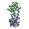
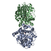
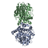
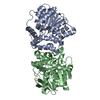
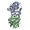
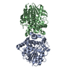
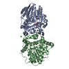
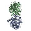
| Title | Fluoroacetate dehalogenase, room temperature structure, using first 1 degree of total 3 degree oscillation | |||||||||
 Components Components | Fluoroacetate dehalogenase | |||||||||
 Keywords Keywords | HYDROLASE / Dehalogenase / defluorinase | |||||||||
| Function / homology |  Function and homology information Function and homology information | |||||||||
| Biological species |  Rhodopseudomonas palustris CGA009 (phototrophic) Rhodopseudomonas palustris CGA009 (phototrophic) | |||||||||
| Method |  X-RAY DIFFRACTION / SYNCHROTRON / MOLECULAR REPLACEMENT / Resolution: 1.8 Å X-RAY DIFFRACTION / SYNCHROTRON / MOLECULAR REPLACEMENT / Resolution: 1.8 Å | |||||||||
 Authors Authors | Finke, A.D. / Wierman, J.L. / Pare-Labrosse, O. / Sarrachini, A. / Besaw, J. / Mehrabi, P. / Gruner, S.M. / Miller, R.J.D. | |||||||||
| Funding support |  United States, 2items United States, 2items
| |||||||||
 Citation Citation | Journal: IUCrJ / Year: 2019 Title: Fixed-target serial oscillation crystallography at room temperature. Authors: Wierman, J.L. / Pare-Labrosse, O. / Sarracini, A. / Besaw, J.E. / Cook, M.J. / Oghbaey, S. / Daoud, H. / Mehrabi, P. / Kriksunov, I. / Kuo, A. / Schuller, D.J. / Smith, S. / Ernst, O.P. / ...Authors: Wierman, J.L. / Pare-Labrosse, O. / Sarracini, A. / Besaw, J.E. / Cook, M.J. / Oghbaey, S. / Daoud, H. / Mehrabi, P. / Kriksunov, I. / Kuo, A. / Schuller, D.J. / Smith, S. / Ernst, O.P. / Szebenyi, D.M.E. / Gruner, S.M. / Miller, R.J.D. / Finke, A.D. | |||||||||
| History |
|
- Structure visualization
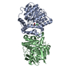
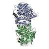
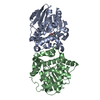
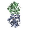
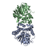
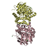
| Structure viewer | Molecule: MolmilJmol/JSmol |
|---|
- Downloads & links
-Download
| PDBx/mmCIF format | 6mzz.cif.gz | 165.4 KB | Display | PDBx/mmCIF format |
|---|---|---|---|---|
| PDB format | pdb6mzz.ent.gz | 104.4 KB | Display | PDB format |
| PDBx/mmJSON format | 6mzz.json.gz | Tree view | PDBx/mmJSON format | |
| Others |  Other downloads Other downloads |
-Validation report
| Arichive directory | https://data.pdbj.org/pub/pdb/validation_reports/mz/6mzzftp://data.pdbj.org/pub/pdb/validation_reports/mz/6mzz | HTTPS FTP |
|---|
-Related structure data
| Related structure data | 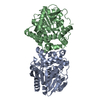 6muhC  6muyC 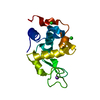 6muzC 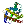 6mv0C 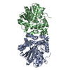 6n00C 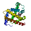 6n02C 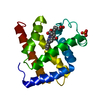 6n03C 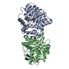 6fsxS C: citing same article ( S: Starting model for refinement |
|---|---|
| Similar structure data |


-Links
 PDBj
PDBj


- Assembly
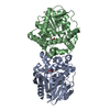
| Deposited unit | 
| ||||||||||||
|---|---|---|---|---|---|---|---|---|---|---|---|---|---|
| 1 |
| ||||||||||||
| Unit cell |
|
-Components
| #1: Protein | Mass: 34073.660 Da / Num. of mol.: 2 Source method: isolated from a genetically manipulated source Source: (gene. exp.) Rhodopseudomonas palustris CGA009 (phototrophic)Strain: ATCC BAA-98 / CGA009 / Gene: RPA1163 / Production host: #2: Chemical | ChemComp-CA / |   Mass: 40.078 Da / Num. of mol.: 1 / Source method: obtained synthetically / Formula: Ca Mass: 40.078 Da / Num. of mol.: 1 / Source method: obtained synthetically / Formula: Ca#3: Water | ChemComp-HOH / |  Mass: 18.015 Da / Num. of mol.: 217 / Source method: isolated from a natural source / Formula: H2O Mass: 18.015 Da / Num. of mol.: 217 / Source method: isolated from a natural source / Formula: H2O |
|---|
-Experimental details
-Experiment
| Experiment | Method: X-RAY DIFFRACTION / Number of used crystals: 1 |
|---|
- Sample preparation
| Crystal | Density Matthews: 1.97 Å3/Da / Density % sol: 37.61 % |
|---|---|
| Crystal grow | Temperature: 295 K / Method: batch mode Details: 16-20% PEG 3350, 100mM Tris-Cl pH 8.5, and 200mM CaCl2. |
-Data collection
| Diffraction | Mean temperature: 298 K / Serial crystal experiment: Y |
|---|---|
| Diffraction source | Source: SYNCHROTRON / Site: CHESS / Beamline: G3 / Wavelength: 1.216 Å |
| Detector | Type: DECTRIS EIGER X 1M / Detector: PIXEL / Date: Mar 1, 2018 |
| Radiation | Monochromator: W/B4C mirrors / Protocol: SINGLE WAVELENGTH / Monochromatic (M) / Laue (L): M / Scattering type: x-ray |
| Radiation wavelength | Wavelength: 1.216 Å / Relative weight: 1 |
| Reflection | Resolution: 1.8→39.55 Å / Num. obs: 48562 / % possible obs: 99.02 % / Redundancy: 5.7 % / Biso Wilson estimate: 34.41 Å2 / CC1/2: 0.954 / Rpim(I) all: 0.1648 / Rrim(I) all: 0.4132 / Net I/σ(I): 4.71 |
| Reflection shell | Resolution: 1.8→1.864 Å / Num. unique obs: 4680 / CC1/2: 0.0739 / Rpim(I) all: 3.646 / Rrim(I) all: 8.37 |
| Serial crystallography measurement | Collimation: Compound refractive lens |
| Serial crystallography sample delivery | Description: Si fixed target / Method: fixed target |
| Serial crystallography data reduction | Crystal hits: 1082 / Frames indexed: 1970 / Frames total: 32000 |
- Processing
| Software |
| ||||||||||||||||||||||||||||||||||||||||||||||||||||||||||||||||||||||||||||||||||||||||||||||||||||||||||||||||||||||||||||||
|---|---|---|---|---|---|---|---|---|---|---|---|---|---|---|---|---|---|---|---|---|---|---|---|---|---|---|---|---|---|---|---|---|---|---|---|---|---|---|---|---|---|---|---|---|---|---|---|---|---|---|---|---|---|---|---|---|---|---|---|---|---|---|---|---|---|---|---|---|---|---|---|---|---|---|---|---|---|---|---|---|---|---|---|---|---|---|---|---|---|---|---|---|---|---|---|---|---|---|---|---|---|---|---|---|---|---|---|---|---|---|---|---|---|---|---|---|---|---|---|---|---|---|---|---|---|---|---|
| Refinement | Method to determine structure: MOLECULAR REPLACEMENT Starting model: 6FSX Resolution: 1.8→39.55 Å / SU ML: 0.3456 / Cross valid method: FREE R-VALUE / σ(F): 1.33 / Phase error: 27.0932
| ||||||||||||||||||||||||||||||||||||||||||||||||||||||||||||||||||||||||||||||||||||||||||||||||||||||||||||||||||||||||||||||
| Solvent computation | Shrinkage radii: 0.9 Å / VDW probe radii: 1.11 Å | ||||||||||||||||||||||||||||||||||||||||||||||||||||||||||||||||||||||||||||||||||||||||||||||||||||||||||||||||||||||||||||||
| Displacement parameters | Biso mean: 38.07 Å2 | ||||||||||||||||||||||||||||||||||||||||||||||||||||||||||||||||||||||||||||||||||||||||||||||||||||||||||||||||||||||||||||||
| Refinement step | Cycle: LAST / Resolution: 1.8→39.55 Å
| ||||||||||||||||||||||||||||||||||||||||||||||||||||||||||||||||||||||||||||||||||||||||||||||||||||||||||||||||||||||||||||||
| Refine LS restraints |
| ||||||||||||||||||||||||||||||||||||||||||||||||||||||||||||||||||||||||||||||||||||||||||||||||||||||||||||||||||||||||||||||
| LS refinement shell |
|
