 Movie
Movie Controller

[English] 日本語
 Yorodumi
- PDB-5k26: Structure of the SH3 domain of MLK3 bound to peptide generated fr... -
+ Open data

- Basic information
| Entry | Database: PDB / ID: 5k26 | ||||||
|---|---|---|---|---|---|---|---|
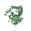
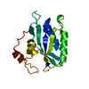
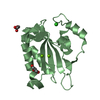
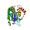
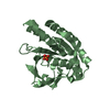
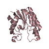
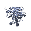
| Title | Structure of the SH3 domain of MLK3 bound to peptide generated from phage display | ||||||
 Components Components | Mitogen-activated protein kinase kinase kinase 11,Chimera protein of MLK3-SH3 and MIP | ||||||
 Keywords Keywords | TRANSFERASE / MLK3 / SH3 / phage display / signalling protein | ||||||
| Function / homology |  Function and homology information Function and homology informationmitogen-activated protein kinase kinase kinase / positive regulation of JUN kinase activity / cell cycle G1/S phase transition / JUN kinase kinase kinase activity / mitogen-activated protein kinase kinase binding / mitogen-activated protein kinase kinase kinase binding / RHOV GTPase cycle / CDC42 GTPase cycle / RHOG GTPase cycle / MAP kinase kinase kinase activity ...mitogen-activated protein kinase kinase kinase / positive regulation of JUN kinase activity / cell cycle G1/S phase transition / JUN kinase kinase kinase activity / mitogen-activated protein kinase kinase binding / mitogen-activated protein kinase kinase kinase binding / RHOV GTPase cycle / CDC42 GTPase cycle / RHOG GTPase cycle / MAP kinase kinase kinase activity / microtubule-based process / JNK cascade / RAF activation / positive regulation of JNK cascade / small GTPase binding / Signaling by moderate kinase activity BRAF mutants / Paradoxical activation of RAF signaling by kinase inactive BRAF / Signaling downstream of RAS mutants / protein autophosphorylation / positive regulation of neuron apoptotic process / MAPK cascade / microtubule / protein phosphorylation / protein kinase activity / protein serine kinase activity / protein serine/threonine kinase activity / centrosome / protein homodimerization activity / ATP binding / membrane / identical protein binding / cytoplasm / cytosol Similarity search - Function | ||||||
| Biological species |  Homo sapiens (human) Homo sapiens (human) | ||||||
| Method |  X-RAY DIFFRACTION / SYNCHROTRON / MOLECULAR REPLACEMENT / Resolution: 1.2 Å X-RAY DIFFRACTION / SYNCHROTRON / MOLECULAR REPLACEMENT / Resolution: 1.2 Å | ||||||
 Authors Authors | Kall, S.K. / Lavie, A. | ||||||
 Citation Citation | Journal: J. Biol. Chem. / Year: 2018 Title: Identification of two distinct peptide-binding pockets in the SH3 domain of human mixed-lineage kinase 3. Authors: Kokoszka, M.E. / Kall, S.L. / Khosla, S. / McGinnis, J.E. / Lavie, A. / Kay, B.K. | ||||||
| History |
|
- Structure visualization
| Structure viewer | Molecule: MolmilJmol/JSmol |
|---|
- Downloads & links
-Download
| PDBx/mmCIF format | 5k26.cif.gz | 82.8 KB | Display | PDBx/mmCIF format |
|---|---|---|---|---|
| PDB format | pdb5k26.ent.gz | 61.7 KB | Display | PDB format |
| PDBx/mmJSON format | 5k26.json.gz | Tree view | PDBx/mmJSON format | |
| Others |  Other downloads Other downloads |
-Validation report
| Arichive directory | https://data.pdbj.org/pub/pdb/validation_reports/k2/5k26ftp://data.pdbj.org/pub/pdb/validation_reports/k2/5k26 | HTTPS FTP |
|---|
-Related structure data
| Related structure data |  5k28C 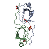 6aqbC  1semS C: citing same article ( S: Starting model for refinement |
|---|---|
| Similar structure data |


-Links
 PDBj
PDBj

- Assembly
| Deposited unit | 
| ||||||||
|---|---|---|---|---|---|---|---|---|---|
| 1 |
| ||||||||
| Unit cell |
|
-Components
| #1: Protein | Mass: 9361.192 Da / Num. of mol.: 2 Fragment: SH3 domain (UNP residues 41-105),SH3 domain (UNP residues 41-105) Source method: isolated from a genetically manipulated source Source: (gene. exp.) Homo sapiens (human) / Gene: MAP3K11, MLK3, PTK1, SPRK / Production host:  References: UniProt: Q16584, mitogen-activated protein kinase kinase kinase #2: Chemical | ChemComp-MES / |   Mass: 195.237 Da / Num. of mol.: 1 / Source method: obtained synthetically / Formula: C6H13NO4S / Comment: pH buffer*YM Mass: 195.237 Da / Num. of mol.: 1 / Source method: obtained synthetically / Formula: C6H13NO4S / Comment: pH buffer*YM#3: Water | ChemComp-HOH / |  Mass: 18.015 Da / Num. of mol.: 123 / Source method: isolated from a natural source / Formula: H2O Mass: 18.015 Da / Num. of mol.: 123 / Source method: isolated from a natural source / Formula: H2O |
|---|
-Experimental details
-Experiment
| Experiment | Method: X-RAY DIFFRACTION / Number of used crystals: 1 |
|---|
- Sample preparation
| Crystal | Density Matthews: 2.31 Å3/Da / Density % sol: 48.53 % |
|---|---|
| Crystal grow | Temperature: 298 K / Method: vapor diffusion, hanging drop / pH: 6.5 Details: 0.1 M Na-Phosphate, 0.1 M K-Phosphate, 0.1 M MES 6.5, 1.5 M NaCl |
-Data collection
| Diffraction | Mean temperature: 100 K |
|---|---|
| Diffraction source | Source: SYNCHROTRON / Site: APS  / Beamline: 21-ID-F / Wavelength: 0.97872 Å / Beamline: 21-ID-F / Wavelength: 0.97872 Å |
| Detector | Type: MARMOSAIC 225 mm CCD / Detector: CCD / Date: Mar 25, 2015 |
| Radiation | Protocol: SINGLE WAVELENGTH / Monochromatic (M) / Laue (L): M / Scattering type: x-ray |
| Radiation wavelength | Wavelength: 0.97872 Å / Relative weight: 1 |
| Reflection | Resolution: 1.2→55.43 Å / Num. obs: 48242 / % possible obs: 97.2 % / Redundancy: 4.92 % / Rmerge(I) obs: 0.042 / Net I/σ(I): 19.53 |
| Reflection shell | Resolution: 1.2→1.27 Å / Redundancy: 2.55 % / Rmerge(I) obs: 0.418 / Mean I/σ(I) obs: 2.76 / % possible all: 83.1 |
- Processing
| Software |
| ||||||||||||||||||||||||||||||||||||||||||||||||||||||||||||||||||||||||||||||||||||||||||||||||||||||||||||||||||||||||||||||||||||||||||||||||||||||||||||||||||||||||||||||||||||||
|---|---|---|---|---|---|---|---|---|---|---|---|---|---|---|---|---|---|---|---|---|---|---|---|---|---|---|---|---|---|---|---|---|---|---|---|---|---|---|---|---|---|---|---|---|---|---|---|---|---|---|---|---|---|---|---|---|---|---|---|---|---|---|---|---|---|---|---|---|---|---|---|---|---|---|---|---|---|---|---|---|---|---|---|---|---|---|---|---|---|---|---|---|---|---|---|---|---|---|---|---|---|---|---|---|---|---|---|---|---|---|---|---|---|---|---|---|---|---|---|---|---|---|---|---|---|---|---|---|---|---|---|---|---|---|---|---|---|---|---|---|---|---|---|---|---|---|---|---|---|---|---|---|---|---|---|---|---|---|---|---|---|---|---|---|---|---|---|---|---|---|---|---|---|---|---|---|---|---|---|---|---|---|---|
| Refinement | Method to determine structure: MOLECULAR REPLACEMENT Starting model: 1SEM Resolution: 1.2→55.43 Å / Cor.coef. Fo:Fc: 0.981 / Cor.coef. Fo:Fc free: 0.973 / SU B: 1.764 / SU ML: 0.033 / Cross valid method: THROUGHOUT / ESU R: 0.034 / ESU R Free: 0.036 / Stereochemistry target values: MAXIMUM LIKELIHOOD / Details: HYDROGENS HAVE BEEN ADDED IN THE RIDING POSITIONS
| ||||||||||||||||||||||||||||||||||||||||||||||||||||||||||||||||||||||||||||||||||||||||||||||||||||||||||||||||||||||||||||||||||||||||||||||||||||||||||||||||||||||||||||||||||||||
| Solvent computation | Ion probe radii: 0.8 Å / Shrinkage radii: 0.8 Å / VDW probe radii: 1.2 Å / Solvent model: MASK | ||||||||||||||||||||||||||||||||||||||||||||||||||||||||||||||||||||||||||||||||||||||||||||||||||||||||||||||||||||||||||||||||||||||||||||||||||||||||||||||||||||||||||||||||||||||
| Displacement parameters | Biso mean: 21.026 Å2
| ||||||||||||||||||||||||||||||||||||||||||||||||||||||||||||||||||||||||||||||||||||||||||||||||||||||||||||||||||||||||||||||||||||||||||||||||||||||||||||||||||||||||||||||||||||||
| Refinement step | Cycle: LAST / Resolution: 1.2→55.43 Å
| ||||||||||||||||||||||||||||||||||||||||||||||||||||||||||||||||||||||||||||||||||||||||||||||||||||||||||||||||||||||||||||||||||||||||||||||||||||||||||||||||||||||||||||||||||||||
| Refine LS restraints |
|