 Movie
Movie Controller


+ Open data

- Basic information
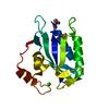
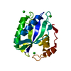
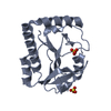
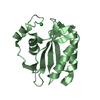
| Entry | Database: PDB / ID: 1hka | ||||||
|---|---|---|---|---|---|---|---|
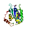
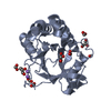
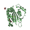
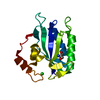
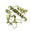
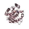
| Title | 6-HYDROXYMETHYL-7,8-DIHYDROPTERIN PYROPHOSPHOKINASE | ||||||
 Components Components | 6-HYDROXYMETHYL-7,8-DIHYDROPTERIN PYROPHOSPHOKINASE | ||||||
 Keywords Keywords | TRANSFERASE / PYROPHOSPHORYL TRANSFER / PYROPHOSPHOKINASE / KINASE / FOLATE / 6-HYDROXYMETHYL-7 / 8-DIHYDROPTERIN | ||||||
| Function / homology |  Function and homology information Function and homology information2-amino-4-hydroxy-6-hydroxymethyldihydropteridine diphosphokinase / 2-amino-4-hydroxy-6-hydroxymethyldihydropteridine diphosphokinase activity / folic acid biosynthetic process / tetrahydrofolate biosynthetic process / kinase activity / magnesium ion binding / ATP binding Similarity search - Function | ||||||
| Biological species |  | ||||||
| Method |  X-RAY DIFFRACTION / SYNCHROTRON / MAD / Resolution: 1.5 Å X-RAY DIFFRACTION / SYNCHROTRON / MAD / Resolution: 1.5 Å | ||||||
 Authors Authors | Xiao, B. / Shi, G. / Chen, X. / Yan, H. / Ji, X. | ||||||
 Citation Citation | Journal: Structure Fold.Des. / Year: 1999 Title: Crystal structure of 6-hydroxymethyl-7,8-dihydropterin pyrophosphokinase, a potential target for the development of novel antimicrobial agents. Authors: Xiao, B. / Shi, G. / Chen, X. / Yan, H. / Ji, X. #1: Journal: J.Bacteriol. / Year: 1992Title: Cloning, Sequence Analysis, and Overexpression of Escherichia Coli Folk, the Gene Coding for 7,8-Dihydro-6-Hydroxymethylpterin-Pyrophosphokinase Authors: Talarico, T.L. / Ray, P.H. / Dev, I.K. / Merrill, B.M. / Dallas, W.S. #2: Journal: J.Bacteriol. / Year: 1991Title: Purification and Partial Characterization of 7,8-Dihydro-6-Hydroxymethylpterin-Pyrophosphokinase and 7,8-Dihydropteroate Synthase from Escherichia Coli Mc4100 Authors: Talarico, T.L. / Dev, I.K. / Dallas, W.S. / Ferone, R. / Ray, P.H. | ||||||
| History |
|
- Structure visualization
| Structure viewer | Molecule: MolmilJmol/JSmol |
|---|
- Downloads & links
-Download
| PDBx/mmCIF format | 1hka.cif.gz | 50.9 KB | Display | PDBx/mmCIF format |
|---|---|---|---|---|
| PDB format | pdb1hka.ent.gz | 35.3 KB | Display | PDB format |
| PDBx/mmJSON format | 1hka.json.gz | Tree view | PDBx/mmJSON format | |
| Others |  Other downloads Other downloads |
-Validation report
| Arichive directory | https://data.pdbj.org/pub/pdb/validation_reports/hk/1hkaftp://data.pdbj.org/pub/pdb/validation_reports/hk/1hka | HTTPS FTP |
|---|
-Related structure data
| Similar structure data |
|---|
-Links
 PDBj
PDBj- Assembly
| Deposited unit | 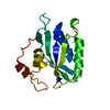
| ||||||||
|---|---|---|---|---|---|---|---|---|---|
| 1 |
| ||||||||
| Unit cell |
|
-Components
| #1: Protein | Mass: 17966.535 Da / Num. of mol.: 1 Source method: isolated from a genetically manipulated source Source: (gene. exp.) References: UniProt: P26281, 2-amino-4-hydroxy-6-hydroxymethyldihydropteridine diphosphokinase |
|---|---|
| #2: Water | ChemComp-HOH /  Mass: 18.015 Da / Num. of mol.: 236 / Source method: isolated from a natural source / Formula: H2O Mass: 18.015 Da / Num. of mol.: 236 / Source method: isolated from a natural source / Formula: H2O |
-Experimental details
-Experiment
| Experiment | Method: X-RAY DIFFRACTION / Number of used crystals: 1 |
|---|
- Sample preparation
| Crystal | Density Matthews: 1.66 Å3/Da / Density % sol: 26 % | ||||||||||||||||||||
|---|---|---|---|---|---|---|---|---|---|---|---|---|---|---|---|---|---|---|---|---|---|
| Crystal grow | pH: 8.5 / Details: pH 8.5 | ||||||||||||||||||||
| Crystal grow | *PLUS Method: vapor diffusion, hanging drop | ||||||||||||||||||||
| Components of the solutions | *PLUS
|
-Data collection
| Diffraction | Mean temperature: 95 K | |||||||||||||||
|---|---|---|---|---|---|---|---|---|---|---|---|---|---|---|---|---|
| Diffraction source | Source: SYNCHROTRON / Site: NSLS  / Beamline: X9B / Wavelength: 0.9686, 0.9788, 0.9792, 0.9870 / Beamline: X9B / Wavelength: 0.9686, 0.9788, 0.9792, 0.9870 | |||||||||||||||
| Detector | Type: MARRESEARCH / Detector: IMAGE PLATE / Date: Aug 20, 1997 / Details: MIRROR | |||||||||||||||
| Radiation | Monochromator: SI(111) / Protocol: MAD / Monochromatic (M) / Laue (L): M / Scattering type: x-ray | |||||||||||||||
| Radiation wavelength |
| |||||||||||||||
| Reflection | Resolution: 1.5→20 Å / Num. obs: 23352 / % possible obs: 95.8 % / Observed criterion σ(I): -0.5 / Redundancy: 5.65 % / Rmerge(I) obs: 0.05 / Net I/σ(I): 21.4 | |||||||||||||||
| Reflection shell | Resolution: 1.5→1.53 Å / Redundancy: 3.81 % / Rmerge(I) obs: 0.34 / Mean I/σ(I) obs: 3.06 / % possible all: 89.9 | |||||||||||||||
| Reflection | *PLUS Num. measured all: 132018 | |||||||||||||||
| Reflection shell | *PLUS % possible obs: 89.9 % |
- Processing
| Software |
| |||||||||||||||||||||||||||||||||
|---|---|---|---|---|---|---|---|---|---|---|---|---|---|---|---|---|---|---|---|---|---|---|---|---|---|---|---|---|---|---|---|---|---|---|
| Refinement | Method to determine structure: MAD / Resolution: 1.5→20 Å / Num. parameters: 6235 / Num. restraintsaints: 5591 / Cross valid method: FREE R IN FIRST STAGE / σ(F): 0 / Stereochemistry target values: ENGH AND HUBER Details: ANISOTROPIC SCALING APPLIED BY THE METHOD OF PARKIN, MOEZZI AND HOPE, J.APPL.CRYST. 28 (1995)53-56
| |||||||||||||||||||||||||||||||||
| Solvent computation | Solvent model: MOEWS & KRETSINGER, J.MOL.BIOL.91(1973)201-2 | |||||||||||||||||||||||||||||||||
| Refine analyze | Num. disordered residues: 8 / Occupancy sum hydrogen: 0 / Occupancy sum non hydrogen: 1503 | |||||||||||||||||||||||||||||||||
| Refinement step | Cycle: LAST / Resolution: 1.5→20 Å
| |||||||||||||||||||||||||||||||||
| Refine LS restraints |
| |||||||||||||||||||||||||||||||||
| Software | *PLUS Name: SHELXL-97 / Classification: refinement | |||||||||||||||||||||||||||||||||
| Refinement | *PLUS σ(I): 2 / Rfactor all: 0.182 / Rfactor obs: 0.174 | |||||||||||||||||||||||||||||||||
| Solvent computation | *PLUS | |||||||||||||||||||||||||||||||||
| Displacement parameters | *PLUS |
