 Movie
Movie Controller

[English] 日本語
 Yorodumi
- PDB-1tf1: Crystal Structure of the E. coli Glyoxylate Regulatory Protein Li... -
+ Open data

- Basic information
| Entry | Database: PDB / ID: 1tf1 | ||||||
|---|---|---|---|---|---|---|---|
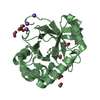
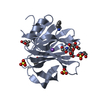
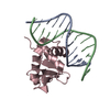
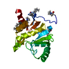
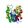
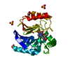
| Title | Crystal Structure of the E. coli Glyoxylate Regulatory Protein Ligand Binding Domain | ||||||
 Components Components | Negative regulator of allantoin and glyoxylate utilization operons | ||||||
 Keywords Keywords | TRANSCRIPTION / Midwest Center for Structural Genomics / GlcR / ligand binding domain / transcriptional regulator / PSI / Protein Structure Initiative / MCSG | ||||||
| Function / homology |  Function and homology information Function and homology informationDNA-binding transcription factor activity / negative regulation of DNA-templated transcription / DNA damage response / DNA-templated transcription / DNA binding / identical protein binding / cytosol Similarity search - Function | ||||||
| Biological species |  | ||||||
| Method |  X-RAY DIFFRACTION / MOLECULAR REPLACEMENT / Resolution: 1.8 Å X-RAY DIFFRACTION / MOLECULAR REPLACEMENT / Resolution: 1.8 Å | ||||||
 Authors Authors | Walker, J.R. / Skarina, T. / Kudrytska, M. / Joachimiak, A. / Arrowsmith, C. / Edwards, A. / Savchenko, A. / Midwest Center for Structural Genomics (MCSG) | ||||||
 Citation Citation | Journal: J.Mol.Biol. / Year: 2006 Title: Structural and biochemical study of effector molecule recognition by the E.coli glyoxylate and allantoin utilization regulatory protein AllR. Authors: Walker, J.R. / Altamentova, S. / Ezersky, A. / Lorca, G. / Skarina, T. / Kudritska, M. / Ball, L.J. / Bochkarev, A. / Savchenko, A. #1: Journal: J.Biol.Chem. / Year: 2002Title: Crystal Structure of Thermotoga maritima 0065, a member of the IclR transcriptional factor family Authors: Zhang, R.G. / Kim, Y. / Skarina, T. / Beasley, S. / Laskowski, R. / Arrowsmith, C. / Edwards, A. / Joachimiak, A. / Savchenko, A. #2: Journal: To be PublishedTitle: Structural Analyses of the Ligand Binding Sites of the IclR family of transcriptional regulators Authors: Walker, J.R. / Evdokimova, L. / Zhang, R.G. / Bochkarev, A. / Joachimiak, A. / Arrowsmith, C. / Edwards, A. / Savchenko, A. | ||||||
| History |
| ||||||
| Remark 300 | BIOMOLECULE GEL FILTRATION STUDIES SHOW THAT THE GLCR LIGAND BINDING DOMAIN FORMS A MONOMER IN SOLUTION. |
- Structure visualization
| Structure viewer | Molecule: MolmilJmol/JSmol |
|---|
- Downloads & links
-Download
| PDBx/mmCIF format | 1tf1.cif.gz | 157.7 KB | Display | PDBx/mmCIF format |
|---|---|---|---|---|
| PDB format | pdb1tf1.ent.gz | 123.2 KB | Display | PDB format |
| PDBx/mmJSON format | 1tf1.json.gz | Tree view | PDBx/mmJSON format | |
| Others |  Other downloads Other downloads |
-Validation report
| Arichive directory | https://data.pdbj.org/pub/pdb/validation_reports/tf/1tf1ftp://data.pdbj.org/pub/pdb/validation_reports/tf/1tf1 | HTTPS FTP |
|---|
-Related structure data
| Related structure data |  1t9l S: Starting model for refinement |
|---|---|
| Similar structure data | |
| Other databases |


-Links
 PDBj
PDBj

- Assembly
| Deposited unit | 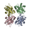
| ||||||||
|---|---|---|---|---|---|---|---|---|---|
| 1 | 
| ||||||||
| 2 | 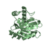
| ||||||||
| 3 | 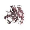
| ||||||||
| 4 | 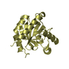
| ||||||||
| Unit cell |
| ||||||||
| Details | The biological assembly is a tetramer generated by applying the translation -46.4850, -48.8411, 0.0000 to molecule C. |
-Components
| #1: Protein | Mass: 21466.574 Da / Num. of mol.: 4 / Fragment: ligand binding domain Source method: isolated from a genetically manipulated source Source: (gene. exp.) #2: Water | ChemComp-HOH / |  Mass: 18.015 Da / Num. of mol.: 595 / Source method: isolated from a natural source / Formula: H2O Mass: 18.015 Da / Num. of mol.: 595 / Source method: isolated from a natural source / Formula: H2O |
|---|
-Experimental details
-Experiment
| Experiment | Method: X-RAY DIFFRACTION / Number of used crystals: 1 |
|---|
- Sample preparation
| Crystal | Density Matthews: 2.18 Å3/Da / Density % sol: 43.56 % |
|---|---|
| Crystal grow | Method: vapor diffusion, hanging drop / pH: 8.5 Details: MgCl2, PEG3350, Tris PH 8.5, ethylene glycol, VAPOR DIFFUSION, HANGING DROP, temperature 100K |
-Data collection
| Diffraction | Mean temperature: 100 K |
|---|---|
| Diffraction source | Source: ROTATING ANODE / Type: RIGAKU RU300 / Wavelength: 1.5418 Å |
| Detector | Type: RIGAKU RAXIS IV / Detector: IMAGE PLATE / Date: Jan 12, 2004 |
| Radiation | Monochromator: CONFOCAL MULTILAYER OPTICS / Protocol: SINGLE WAVELENGTH / Monochromatic (M) / Laue (L): M / Scattering type: x-ray |
| Radiation wavelength | Wavelength: 1.5418 Å / Relative weight: 1 |
| Reflection | Resolution: 1.8→20 Å / Num. all: 59628 / Num. obs: 59628 / % possible obs: 97.9 % / Observed criterion σ(F): 0 / Observed criterion σ(I): -3 / Redundancy: 3 % / Biso Wilson estimate: 20.7 Å2 / Rmerge(I) obs: 0.087 / Net I/σ(I): 15.1 |
| Reflection shell | Resolution: 1.8→1.86 Å / Rmerge(I) obs: 0.277 / Num. unique all: 5986 / % possible all: 98.8 |
- Processing
| Software |
| ||||||||||||||||||||||||||||||||||||
|---|---|---|---|---|---|---|---|---|---|---|---|---|---|---|---|---|---|---|---|---|---|---|---|---|---|---|---|---|---|---|---|---|---|---|---|---|---|
| Refinement | Method to determine structure: MOLECULAR REPLACEMENT Starting model: PDB ENTRY 1T9L 1t9l Resolution: 1.8→19.73 Å / Rfactor Rfree error: 0.005 / Data cutoff high absF: 2980696.7 / Data cutoff low absF: 0 / Isotropic thermal model: RESTRAINED / Cross valid method: THROUGHOUT / σ(F): 0 / Stereochemistry target values: Engh & Huber
| ||||||||||||||||||||||||||||||||||||
| Solvent computation | Solvent model: FLAT MODEL / Bsol: 41.8659 Å2 / ksol: 0.363963 e/Å3 | ||||||||||||||||||||||||||||||||||||
| Displacement parameters | Biso mean: 27.6 Å2
| ||||||||||||||||||||||||||||||||||||
| Refine analyze |
| ||||||||||||||||||||||||||||||||||||
| Refinement step | Cycle: LAST / Resolution: 1.8→19.73 Å
| ||||||||||||||||||||||||||||||||||||
| Refine LS restraints |
| ||||||||||||||||||||||||||||||||||||
| LS refinement shell | Resolution: 1.8→1.91 Å / Rfactor Rfree error: 0.012 / Total num. of bins used: 6
| ||||||||||||||||||||||||||||||||||||
| Xplor file |
|
