sequence-specific double-stranded DNA binding / sequence-specific DNA binding / DNA-binding transcription factor activity, RNA polymerase II-specific / cell differentiation / positive regulation of apoptotic process / regulation of transcription by RNA polymerase II / chromatin / negative regulation of transcription by RNA polymerase II / DNA-templated transcription / nucleus Similarity search - Function
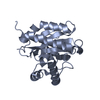
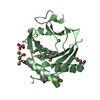
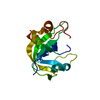
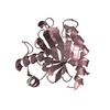
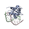
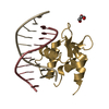
A: Enhancer site of Prostate Specific Antigen Promoter Region B: Enhancer site of Prostate Specific Antigen Promoter Region C: SAM pointed domain containing ets transcription factor
Mass: 4046.645 Da / Num. of mol.: 1 / Fragment: Sense Strand of E site / Source method: obtained synthetically Details: This sequence occurs naturally in humans. DNA substrate for protein is synthesized chemically and purified through chromatography
Mass: 3895.563 Da / Num. of mol.: 1 / Fragment: Antisense Strand of E site / Source method: obtained synthetically Details: This sequence occurs naturally in humans. DNA substrate for protein is synthesized chemically and purified through chromatography
#3: Protein
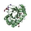
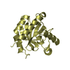
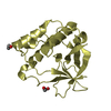
SAMpointeddomaincontainingetstranscriptionfactor
Mass: 11587.606 Da / Num. of mol.: 1 / Fragment: PDEF C-terminal Ets domain Source method: isolated from a genetically manipulated source Source: (gene. exp.) Homo sapiens (human) / Gene: PDEF / Plasmid: PET-GST-T-A / Production host: Escherichia coli (E. coli) / Strain (production host): BL21 (Lambda DE3) / References: UniProt: O95238
In the structure databanks used in Yorodumi, some data are registered as the other names, "COVID-19 virus" and "2019-nCoV". Here are the details of the virus and the list of structure data.
Jan 31, 2019. EMDB accession codes are about to change! (news from PDBe EMDB page)
EMDB accession codes are about to change! (news from PDBe EMDB page)
The allocation of 4 digits for EMDB accession codes will soon come to an end. Whilst these codes will remain in use, new EMDB accession codes will include an additional digit and will expand incrementally as the available range of codes is exhausted. The current 4-digit format prefixed with “EMD-” (i.e. EMD-XXXX) will advance to a 5-digit format (i.e. EMD-XXXXX), and so on. It is currently estimated that the 4-digit codes will be depleted around Spring 2019, at which point the 5-digit format will come into force.
The EM Navigator/Yorodumi systems omit the EMD- prefix.
Related info.:Q: What is EMD? / ID/Accession-code notation in Yorodumi/EM Navigator
Yorodumi is a browser for structure data from EMDB, PDB, SASBDB, etc.
This page is also the successor to EM Navigator detail page, and also detail information page/front-end page for Omokage search.
The word "yorodu" (or yorozu) is an old Japanese word meaning "ten thousand". "mi" (miru) is to see.
Related info.:EMDB / PDB / SASBDB / Comparison of 3 databanks / Yorodumi Search / Aug 31, 2016. New EM Navigator & Yorodumi / Yorodumi Papers / Jmol/JSmol / Function and homology information / Changes in new EM Navigator and Yorodumi
 Movie
Movie Controller

 Yorodumi
 Open data
 Basic information
 Components
 Keywords
 Function and homology information
 Homo sapiens (human)
 X-RAY DIFFRACTION /
  Authors
 Citation
 Structure visualization
 Downloads & links
 Other downloads

 PDBj
PDBj


 Assembly
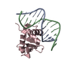

 Mass: 18.015 Da / Num. of mol.: 156 / Source method: isolated from a natural source / Formula: H2O
 Sample preparation
 Processing