 Movie
Movie Controller

[English] 日本語
 Yorodumi
- PDB-2p5d: Crystal structure of MJECL36 from Methanocaldococcus jannaschii D... -
+ Open data

- Basic information
| Entry | Database: PDB / ID: 2p5d | ||||||
|---|---|---|---|---|---|---|---|
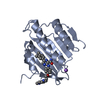
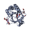
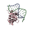
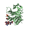
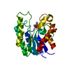
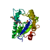
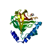
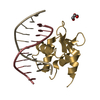
| Title | Crystal structure of MJECL36 from Methanocaldococcus jannaschii DSM 2661 | ||||||
 Components Components | UPF0310 protein MJECL36 | ||||||
 Keywords Keywords | STRUCTURAL GENOMICS / UNKNOWN FUNCTION / Methanocaldococcus jannaschii / NPPSFA / National Project on Protein Structural and Functional Analyses / RIKEN Structural Genomics/Proteomics Initiative / RSGI | ||||||
| Function / homology | Uncharacterised protein family UPF0310 / EVE domain / EVE domain / ph1033 like fold / ph1033 like domains / PUA-like superfamily / Roll / Alpha Beta / UPF0310 protein MJECL36 Function and homology information Function and homology information | ||||||
| Biological species |   Methanocaldococcus jannaschii (archaea) Methanocaldococcus jannaschii (archaea) | ||||||
| Method |  X-RAY DIFFRACTION / MOLECULAR REPLACEMENT / Resolution: 1.7 Å X-RAY DIFFRACTION / MOLECULAR REPLACEMENT / Resolution: 1.7 Å | ||||||
 Authors Authors | Sugahara, M. / Kunishima, N. / RIKEN Structural Genomics/Proteomics Initiative (RSGI) | ||||||
 Citation Citation | Journal: To be Published Title: Crystal structure of MJECL36 from Methanocaldococcus jannaschii DSM 2661 Authors: Sugahara, M. / Kunishima, N. | ||||||
| History |
|
- Structure visualization
| Structure viewer | Molecule: MolmilJmol/JSmol |
|---|
- Downloads & links
-Download
| PDBx/mmCIF format | 2p5d.cif.gz | 47.6 KB | Display | PDBx/mmCIF format |
|---|---|---|---|---|
| PDB format | pdb2p5d.ent.gz | 32.6 KB | Display | PDB format |
| PDBx/mmJSON format | 2p5d.json.gz | Tree view | PDBx/mmJSON format | |
| Others |  Other downloads Other downloads |
-Validation report
| Arichive directory | https://data.pdbj.org/pub/pdb/validation_reports/p5/2p5dftp://data.pdbj.org/pub/pdb/validation_reports/p5/2p5d | HTTPS FTP |
|---|
-Related structure data
| Related structure data | 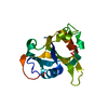 1wmmS S: Starting model for refinement |
|---|---|
| Similar structure data | |
| Other databases |


-Links
 PDBj
PDBj
- Assembly
| Deposited unit | 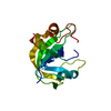
| ||||||||
|---|---|---|---|---|---|---|---|---|---|
| 1 |
| ||||||||
| Unit cell |
| ||||||||
| Details | The biological assembly is monomer in the asymmetric. |
-Components
| #1: Protein | Mass: 17574.719 Da / Num. of mol.: 1 Source method: isolated from a genetically manipulated source Source: (gene. exp.) Methanocaldococcus jannaschii (archaea)Plasmid: pET-21a / Production host:  |
|---|---|
| #2: Water | ChemComp-HOH /  Mass: 18.015 Da / Num. of mol.: 194 / Source method: isolated from a natural source / Formula: H2O Mass: 18.015 Da / Num. of mol.: 194 / Source method: isolated from a natural source / Formula: H2O |
-Experimental details
-Experiment
| Experiment | Method: X-RAY DIFFRACTION / Number of used crystals: 1 |
|---|
- Sample preparation
| Crystal | Density Matthews: 2.05 Å3/Da / Density % sol: 39.88 % |
|---|---|
| Crystal grow | Temperature: 291 K / Method: oil microbatch / pH: 5.5 Details: 27.5% PEG 4000, 0.1M Acetate-NaOH, 10% Dioxane, pH 5.5, oil microbatch, temperature 291K |
-Data collection
| Diffraction | Mean temperature: 100 K |
|---|---|
| Diffraction source | Source: ROTATING ANODE / Type: RIGAKU / Wavelength: 1.54 Å |
| Detector | Type: RIGAKU RAXIS VII / Detector: IMAGE PLATE / Date: Jan 25, 2007 |
| Radiation | Protocol: SINGLE WAVELENGTH / Monochromatic (M) / Laue (L): M / Scattering type: x-ray |
| Radiation wavelength | Wavelength: 1.54 Å / Relative weight: 1 |
| Reflection | Resolution: 1.7→20 Å / Num. all: 16540 / Num. obs: 16540 / % possible obs: 100 % / Observed criterion σ(F): 0 / Observed criterion σ(I): 0 / Redundancy: 6.7 % / Biso Wilson estimate: 19.38 Å2 / Rmerge(I) obs: 0.04 / Rsym value: 0.037 / Net I/σ(I): 15.3 |
| Reflection shell | Resolution: 1.7→1.76 Å / Redundancy: 6.5 % / Rmerge(I) obs: 0.189 / Mean I/σ(I) obs: 6.9 / Num. unique all: 1625 / Rsym value: 0.176 / % possible all: 100 |
- Processing
| Software |
| |||||||||||||||||||||||||
|---|---|---|---|---|---|---|---|---|---|---|---|---|---|---|---|---|---|---|---|---|---|---|---|---|---|---|
| Refinement | Method to determine structure: MOLECULAR REPLACEMENT Starting model: PDB ENTRY 1WMM Resolution: 1.7→19.25 Å / Isotropic thermal model: Anisotrop / Cross valid method: THROUGHOUT / σ(F): 0 / Stereochemistry target values: Engh & Huber
| |||||||||||||||||||||||||
| Displacement parameters | Biso mean: 21.3 Å2
| |||||||||||||||||||||||||
| Refine analyze |
| |||||||||||||||||||||||||
| Refinement step | Cycle: LAST / Resolution: 1.7→19.25 Å
| |||||||||||||||||||||||||
| Refine LS restraints |
| |||||||||||||||||||||||||
| LS refinement shell | Resolution: 1.7→1.76 Å / Rfactor Rfree error: 0.03
|
