 Movie
Movie Controller

[English] 日本語
 Yorodumi
- PDB-5jvt: Crystal structure of the DNA binding domain of transcription fact... -
+ Open data

- Basic information
| Entry | Database: PDB / ID: 5jvt | ||||||
|---|---|---|---|---|---|---|---|
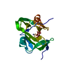
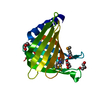
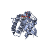
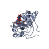
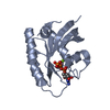
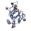
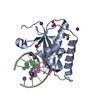
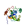
| Title | Crystal structure of the DNA binding domain of transcription factor FLI1 in complex with an 11-mer DNA GACCGGAAGTG | ||||||
 Components Components |
| ||||||
 Keywords Keywords | transcription/dna / transcription / DNA binding / cancer / Ewing sarcoma / transcription-dna complex | ||||||
| Function / homology |  Function and homology information Function and homology informationhemostasis / animal organ morphogenesis / Transcriptional regulation of granulopoiesis / sequence-specific double-stranded DNA binding / DNA-binding transcription activator activity, RNA polymerase II-specific / DNA-binding transcription factor activity, RNA polymerase II-specific / nuclear body / cell differentiation / transcription cis-regulatory region binding / RNA polymerase II cis-regulatory region sequence-specific DNA binding ...hemostasis / animal organ morphogenesis / Transcriptional regulation of granulopoiesis / sequence-specific double-stranded DNA binding / DNA-binding transcription activator activity, RNA polymerase II-specific / DNA-binding transcription factor activity, RNA polymerase II-specific / nuclear body / cell differentiation / transcription cis-regulatory region binding / RNA polymerase II cis-regulatory region sequence-specific DNA binding / DNA-binding transcription factor activity / chromatin binding / regulation of transcription by RNA polymerase II / positive regulation of DNA-templated transcription / chromatin / DNA-templated transcription / DNA binding / nucleoplasm / nucleus Similarity search - Function | ||||||
| Biological species |  Homo sapiens (human) Homo sapiens (human) Endothia gyrosa (fungus) Endothia gyrosa (fungus) | ||||||
| Method |  X-RAY DIFFRACTION / SYNCHROTRON / MOLECULAR REPLACEMENT / Resolution: 3.1 Å X-RAY DIFFRACTION / SYNCHROTRON / MOLECULAR REPLACEMENT / Resolution: 3.1 Å | ||||||
 Authors Authors | Hou, C. / Tsodikov, O.V. | ||||||
 Citation Citation | Journal: Nucleic Acids Res. / Year: 2016 Title: Structures of mithramycin analogues bound to DNA and implications for targeting transcription factor FLI1. Authors: Hou, C. / Weidenbach, S. / Cano, K.E. / Wang, Z. / Mitra, P. / Ivanov, D.N. / Rohr, J. / Tsodikov, O.V. | ||||||
| History |
|
- Structure visualization
| Structure viewer | Molecule: MolmilJmol/JSmol |
|---|
- Downloads & links
-Download
| PDBx/mmCIF format | 5jvt.cif.gz | 206.5 KB | Display | PDBx/mmCIF format |
|---|---|---|---|---|
| PDB format | pdb5jvt.ent.gz | 162.1 KB | Display | PDB format |
| PDBx/mmJSON format | 5jvt.json.gz | Tree view | PDBx/mmJSON format | |
| Others |  Other downloads Other downloads |
-Validation report
| Arichive directory | https://data.pdbj.org/pub/pdb/validation_reports/jv/5jvtftp://data.pdbj.org/pub/pdb/validation_reports/jv/5jvt | HTTPS FTP |
|---|
-Related structure data
| Related structure data |  5jvwC 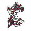 5jw0C  5jw2C  5e8iS C: citing same article ( S: Starting model for refinement |
|---|---|
| Similar structure data |


-Links
 PDBj
PDBj


- Assembly
| Deposited unit | 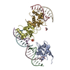
| ||||||||
|---|---|---|---|---|---|---|---|---|---|
| 1 | 
| ||||||||
| 2 | 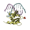
| ||||||||
| 3 | 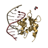
| ||||||||
| Unit cell |
|
-Components
-Protein , 1 types, 3 molecules ADG
| #1: Protein | Mass: 12098.655 Da / Num. of mol.: 3 Source method: isolated from a genetically manipulated source Source: (gene. exp.) Homo sapiens (human) / Gene: FLI1 / Production host:  |
|---|
-DNA chain , 2 types, 6 molecules BEHCFI
| #2: DNA chain | Mass: 3423.248 Da / Num. of mol.: 3 / Source method: obtained synthetically / Source: (synth.) Endothia gyrosa (fungus)#3: DNA chain | Mass: 3285.148 Da / Num. of mol.: 3 / Source method: obtained synthetically / Source: (synth.) Endothia gyrosa (fungus) |
|---|
-Non-polymers , 3 types, 4 molecules 


| #4: Chemical | ChemComp-PO4 /  Mass: 94.971 Da / Num. of mol.: 1 / Source method: obtained synthetically / Formula: PO4 Mass: 94.971 Da / Num. of mol.: 1 / Source method: obtained synthetically / Formula: PO4 |
|---|---|
| #5: Chemical | ChemComp-GOL /  Mass: 92.094 Da / Num. of mol.: 1 / Source method: obtained synthetically / Formula: C3H8O3 Mass: 92.094 Da / Num. of mol.: 1 / Source method: obtained synthetically / Formula: C3H8O3 |
| #6: Water | ChemComp-HOH / Mass: 18.015 Da / Num. of mol.: 2 / Source method: isolated from a natural source / Formula: H2O |
-Experimental details
-Experiment
| Experiment | Method: X-RAY DIFFRACTION / Number of used crystals: 1 |
|---|
- Sample preparation
| Crystal | Density Matthews: 4.12 Å3/Da / Density % sol: 70.16 % |
|---|---|
| Crystal grow | Temperature: 294 K / Method: vapor diffusion, hanging drop / pH: 4.6 Details: 1.6 M ammonium sulphate, 0.1 M sodium acetate pH 4.6 |
-Data collection
| Diffraction | Mean temperature: 100 K |
|---|---|
| Diffraction source | Source: SYNCHROTRON / Site: APS  / Beamline: 22-ID / Wavelength: 1 Å / Beamline: 22-ID / Wavelength: 1 Å |
| Detector | Type: RAYONIX MX300-HS / Detector: CCD / Date: Jun 4, 2015 |
| Radiation | Protocol: SINGLE WAVELENGTH / Monochromatic (M) / Laue (L): M / Scattering type: x-ray |
| Radiation wavelength | Wavelength: 1 Å / Relative weight: 1 |
| Reflection | Resolution: 3.1→50 Å / Num. obs: 17757 / % possible obs: 99.8 % / Observed criterion σ(I): 2 / Redundancy: 5.9 % / Rmerge(I) obs: 0.11 / Net I/σ(I): 19 |
- Processing
| Software |
| ||||||||||||||||||||||||||||||||||||||||||||||||||||||||||||||||||||||||||||||||||||||||||||||||||||||||||||||||||||||||||||||||||||||||||||||||||||||||||||||||||||||||||||||||||||||
|---|---|---|---|---|---|---|---|---|---|---|---|---|---|---|---|---|---|---|---|---|---|---|---|---|---|---|---|---|---|---|---|---|---|---|---|---|---|---|---|---|---|---|---|---|---|---|---|---|---|---|---|---|---|---|---|---|---|---|---|---|---|---|---|---|---|---|---|---|---|---|---|---|---|---|---|---|---|---|---|---|---|---|---|---|---|---|---|---|---|---|---|---|---|---|---|---|---|---|---|---|---|---|---|---|---|---|---|---|---|---|---|---|---|---|---|---|---|---|---|---|---|---|---|---|---|---|---|---|---|---|---|---|---|---|---|---|---|---|---|---|---|---|---|---|---|---|---|---|---|---|---|---|---|---|---|---|---|---|---|---|---|---|---|---|---|---|---|---|---|---|---|---|---|---|---|---|---|---|---|---|---|---|---|
| Refinement | Method to determine structure: MOLECULAR REPLACEMENT Starting model: 5E8I Resolution: 3.1→40 Å / Cor.coef. Fo:Fc: 0.94 / Cor.coef. Fo:Fc free: 0.933 / SU ML: 0.323 / Cross valid method: THROUGHOUT / ESU R: 0.814 / ESU R Free: 0.35 / Details: HYDROGENS HAVE BEEN ADDED IN THE RIDING POSITIONS
| ||||||||||||||||||||||||||||||||||||||||||||||||||||||||||||||||||||||||||||||||||||||||||||||||||||||||||||||||||||||||||||||||||||||||||||||||||||||||||||||||||||||||||||||||||||||
| Solvent computation | Ion probe radii: 0.8 Å / Shrinkage radii: 0.8 Å / VDW probe radii: 1.2 Å | ||||||||||||||||||||||||||||||||||||||||||||||||||||||||||||||||||||||||||||||||||||||||||||||||||||||||||||||||||||||||||||||||||||||||||||||||||||||||||||||||||||||||||||||||||||||
| Displacement parameters |
| ||||||||||||||||||||||||||||||||||||||||||||||||||||||||||||||||||||||||||||||||||||||||||||||||||||||||||||||||||||||||||||||||||||||||||||||||||||||||||||||||||||||||||||||||||||||
| Refinement step | Cycle: 1 / Resolution: 3.1→40 Å
| ||||||||||||||||||||||||||||||||||||||||||||||||||||||||||||||||||||||||||||||||||||||||||||||||||||||||||||||||||||||||||||||||||||||||||||||||||||||||||||||||||||||||||||||||||||||
| Refine LS restraints |
|