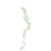
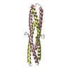
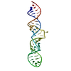
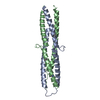
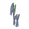
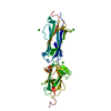
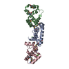
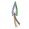
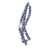
ジャーナル: J Biol Chem / 年: 2016 タイトル: The Structure of the Plakin Domain of Plectin Reveals an Extended Rod-like Shape. 著者: Esther Ortega / José A Manso / Rubén M Buey / Ana M Carballido / Arturo Carabias / Arnoud Sonnenberg / José M de Pereda / 要旨: Plakins are large multi-domain proteins that interconnect cytoskeletal structures. Plectin is a prototypical plakin that tethers intermediate filaments to membrane-associated complexes. Most plakins ...Plakins are large multi-domain proteins that interconnect cytoskeletal structures. Plectin is a prototypical plakin that tethers intermediate filaments to membrane-associated complexes. Most plakins contain a plakin domain formed by up to nine spectrin repeats (SR1-SR9) and an SH3 domain. The plakin domains of plectin and other plakins harbor binding sites for junctional proteins. We have combined x-ray crystallography with small angle x-ray scattering (SAXS) to elucidate the structure of the plakin domain of plectin, extending our previous analysis of the SR1 to SR5 region. Two crystal structures of the SR5-SR6 region allowed us to characterize its uniquely wide inter-repeat conformational variability. We also report the crystal structures of the SR7-SR8 region, refined to 1.8 Å, and the SR7-SR9 at lower resolution. The SR7-SR9 region, which is conserved in all other plakin domains, forms a rigid segment stabilized by uniquely extensive inter-repeat contacts mediated by unusually long helices in SR8 and SR9. Using SAXS we show that in solution the SR3-SR6 and SR7-SR9 regions are rod-like segments and that SR3-SR9 of plectin has an extended shape with a small central kink. Other plakins, such as bullous pemphigoid antigen 1 and microtubule and actin cross-linking factor 1, are likely to have similar extended plakin domains. In contrast, desmoplakin has a two-segment structure with a central flexible hinge. The continuous versus segmented structures of the plakin domains of plectin and desmoplakin give insight into how different plakins might respond to tension and transmit mechanical signals.
分子量: 22323.803 Da / 分子数: 2 / 変異: F752A,F752A / 由来タイプ: 組換発現 詳細: The SH3 domain, residues 819-888, has been replaced by the short sequence GSG 由来: (組換発現) Homo sapiens (ヒト) / 遺伝子: PLEC, PLEC1 / プラスミド: MODIFIED pET15b / 発現宿主: Escherichia coli BL21(DE3) (大腸菌) / 参照: UniProt: Q15149
-
実験情報
-
実験
実験
手法: X線回折 / 使用した結晶の数: 1
-
試料調製
結晶
マシュー密度: 2.97 Å3/Da / 溶媒含有率: 58 %
結晶化
温度: 277 K / 手法: 蒸気拡散法, ハンギングドロップ法 / pH: 6 詳細: A protein solution at 30 mg/ml in 10 mM Tris-Hcl (pH 7.5), 50 mM NaCl, 1 mM DTT was mixed with an equal volume of crystallization solution 0.1 M Bis-Tris-Propane (pH 6.0), 18% PEG 3350, 0.2 Na malonate
-
データ収集
回折
平均測定温度: 100 K
放射光源
由来: 回転陽極 / タイプ: BRUKER AXS MICROSTAR-H / 波長: 1.5418 Å
検出器
タイプ: MAR scanner 345 mm plate / 検出器: IMAGE PLATE / 日付: 2010年10月8日
放射
モノクロメーター: HELIOS OPTICS / プロトコル: SINGLE WAVELENGTH / 単色(M)・ラウエ(L): M / 散乱光タイプ: x-ray
放射波長
波長: 1.5418 Å / 相対比: 1
反射
解像度: 3→43.4 Å / Num. obs: 10510 / % possible obs: 97.7 % / Observed criterion σ(I): -3 / 冗長度: 3.9 % / Biso Wilson estimate: 66.8 Å2 / CC1/2: 0.998 / Rpim(I) all: 0.093 / Net I/σ(I): 15
反射 シェル
解像度: 3→3.08 Å / 冗長度: 3.9 % / Mean I/σ(I) obs: 2 / Rpim(I) all: 0.889 / % possible all: 100
 ムービー
ムービー コントローラー


 データを開く
 基本情報
基本情報 要素
要素 キーワード
キーワード 機能・相同性情報
 Homo sapiens (ヒト)
 X線回折 /
  データ登録者
 スペイン, 1件
  引用
引用
 構造の表示
構造の表示 ダウンロードとリンク
 その他のダウンロード


 PDBj
PDBj


 集合体
集合体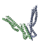


 試料調製
試料調製 解析
解析