 Movie
Movie Controller


+ Open data

- Basic information
| Entry | Database: PDB / ID: 5is2 | ||||||
|---|---|---|---|---|---|---|---|
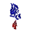
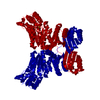
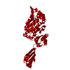
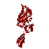
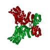
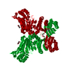
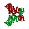
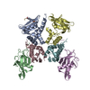
| Title | Crystal structure of Mycobacterium avium SerB2 at pH 6.6 | ||||||
 Components Components | Phosphoserine phosphatase | ||||||
 Keywords Keywords | HYDROLASE / HAD family / phosphoserine phosphatase | ||||||
| Function / homology |  Function and homology information Function and homology informationphosphoserine phosphatase / L-phosphoserine phosphatase activity / L-serine biosynthetic process / magnesium ion binding / cytoplasm Similarity search - Function | ||||||
| Biological species |  Mycobacterium avium (bacteria) Mycobacterium avium (bacteria) | ||||||
| Method |  X-RAY DIFFRACTION / SYNCHROTRON / MOLECULAR REPLACEMENT / Resolution: 1.881 Å X-RAY DIFFRACTION / SYNCHROTRON / MOLECULAR REPLACEMENT / Resolution: 1.881 Å | ||||||
 Authors Authors | Shree, S. / Ramachandran, R. | ||||||
| Funding support |  India, 1items India, 1items
| ||||||
 Citation Citation | Journal: To be published Title: Crystal Structure of Mycobacterium avium SerB2 (MAV_3907) at pH 6.6 Authors: Shree, S. / Ramachandran, R. #1: Journal: J. Struct. Funct. Genomics / Year: 2011Title: SAD phasing using iodide ions in a high-throughput structural genomics environment. Authors: Abendroth, J. / Gardberg, A.S. / Robinson, J.I. / Christensen, J.S. / Staker, B.L. / Myler, P.J. / Stewart, L.J. / Edwards, T.E. | ||||||
| History |
|
- Structure visualization
| Structure viewer | Molecule: MolmilJmol/JSmol |
|---|
- Downloads & links
-Download
| PDBx/mmCIF format | 5is2.cif.gz | 163.8 KB | Display | PDBx/mmCIF format |
|---|---|---|---|---|
| PDB format | pdb5is2.ent.gz | 129 KB | Display | PDB format |
| PDBx/mmJSON format | 5is2.json.gz | Tree view | PDBx/mmJSON format | |
| Others |  Other downloads Other downloads |
-Validation report
| Summary document | 5is2_validation.pdf.gz | 424.9 KB | Display | wwPDB validaton report |
|---|---|---|---|---|
| Full document | 5is2_full_validation.pdf.gz | 427.4 KB | Display | |
| Data in XML | 5is2_validation.xml.gz | 16.1 KB | Display | |
| Data in CIF | 5is2_validation.cif.gz | 22.7 KB | Display | |
| Arichive directory | https://data.pdbj.org/pub/pdb/validation_reports/is/5is2ftp://data.pdbj.org/pub/pdb/validation_reports/is/5is2 | HTTPS FTP |
-Related structure data
| Related structure data |  5jjbC  5t41C  3p96S S: Starting model for refinement C: citing same article ( |
|---|---|
| Similar structure data |


-Links
 PDBj
PDBj

- Assembly
| Deposited unit | 
| ||||||||
|---|---|---|---|---|---|---|---|---|---|
| 1 | 
| ||||||||
| Unit cell |
|
-Components
| #1: Protein | Mass: 41787.820 Da / Num. of mol.: 1 / Fragment: UNP RESIDUES 5-400 / Mutation: G31R Source method: isolated from a genetically manipulated source Source: (gene. exp.) Mycobacterium avium (strain 104) (bacteria)Strain: 104 / Gene: serB, MAV_3907 / Plasmid: pAVA0421 / Production host: |
|---|---|
| #2: Chemical | ChemComp-MG /   Mass: 24.305 Da / Num. of mol.: 1 / Source method: obtained synthetically / Formula: Mg Mass: 24.305 Da / Num. of mol.: 1 / Source method: obtained synthetically / Formula: Mg |
| #3: Water | ChemComp-HOH /  Mass: 18.015 Da / Num. of mol.: 123 / Source method: isolated from a natural source / Formula: H2O Mass: 18.015 Da / Num. of mol.: 123 / Source method: isolated from a natural source / Formula: H2O |
-Experimental details
-Experiment
| Experiment | Method: X-RAY DIFFRACTION / Number of used crystals: 1 |
|---|
- Sample preparation
| Crystal | Density Matthews: 2.9 Å3/Da / Density % sol: 57.58 % Description: Mycobacterium avium SerB2 protein was crystallized in presence of its inhibitor rosaniline (a pink color compound) and pink coloured crystals were diffracted, however no density of ...Description: Mycobacterium avium SerB2 protein was crystallized in presence of its inhibitor rosaniline (a pink color compound) and pink coloured crystals were diffracted, however no density of rosaniline was found on solving the crystal data. |
|---|---|
| Crystal grow | Temperature: 293 K / Method: vapor diffusion, hanging drop / pH: 6.6 Details: 22% PEG 8000, 0.1M Magnesium acetate tetrahydrate, 0.1M HEPES pH 6.6 |
-Data collection
| Diffraction | Mean temperature: 100 K |
|---|---|
| Diffraction source | Source: SYNCHROTRON / Site: ESRF  / Beamline: BM14 / Wavelength: 0.978 Å / Beamline: BM14 / Wavelength: 0.978 Å |
| Detector | Type: MARMOSAIC 225 mm CCD / Detector: CCD / Date: Dec 3, 2013 |
| Radiation | Protocol: SINGLE WAVELENGTH / Monochromatic (M) / Laue (L): M / Scattering type: x-ray |
| Radiation wavelength | Wavelength: 0.978 Å / Relative weight: 1 |
| Reflection | Resolution: 1.88→50 Å / Num. obs: 38932 / % possible obs: 98.4 % / Redundancy: 7.8 % / Net I/σ(I): 29 |
- Processing
| Software |
| |||||||||||||||||||||||||||||||||||||||||||||||||||||||||||||||||||||||||||||||||||||||||||||||||||||||||
|---|---|---|---|---|---|---|---|---|---|---|---|---|---|---|---|---|---|---|---|---|---|---|---|---|---|---|---|---|---|---|---|---|---|---|---|---|---|---|---|---|---|---|---|---|---|---|---|---|---|---|---|---|---|---|---|---|---|---|---|---|---|---|---|---|---|---|---|---|---|---|---|---|---|---|---|---|---|---|---|---|---|---|---|---|---|---|---|---|---|---|---|---|---|---|---|---|---|---|---|---|---|---|---|---|---|---|
| Refinement | Method to determine structure: MOLECULAR REPLACEMENT Starting model: 3P96 Resolution: 1.881→26.139 Å / SU ML: 0.22 / Cross valid method: NONE / σ(F): 1.34 / Phase error: 28.81 / Stereochemistry target values: ML
| |||||||||||||||||||||||||||||||||||||||||||||||||||||||||||||||||||||||||||||||||||||||||||||||||||||||||
| Solvent computation | Shrinkage radii: 0.9 Å / VDW probe radii: 1.11 Å / Solvent model: FLAT BULK SOLVENT MODEL | |||||||||||||||||||||||||||||||||||||||||||||||||||||||||||||||||||||||||||||||||||||||||||||||||||||||||
| Displacement parameters | Biso max: 132.75 Å2 / Biso mean: 53.9969 Å2 / Biso min: 28.59 Å2 | |||||||||||||||||||||||||||||||||||||||||||||||||||||||||||||||||||||||||||||||||||||||||||||||||||||||||
| Refinement step | Cycle: final / Resolution: 1.881→26.139 Å
| |||||||||||||||||||||||||||||||||||||||||||||||||||||||||||||||||||||||||||||||||||||||||||||||||||||||||
| Refine LS restraints |
| |||||||||||||||||||||||||||||||||||||||||||||||||||||||||||||||||||||||||||||||||||||||||||||||||||||||||
| LS refinement shell | Refine-ID: X-RAY DIFFRACTION / Total num. of bins used: 14
| |||||||||||||||||||||||||||||||||||||||||||||||||||||||||||||||||||||||||||||||||||||||||||||||||||||||||
| Refinement TLS params. | Method: refined / Origin x: -4.9413 Å / Origin y: -10.7168 Å / Origin z: -38.6808 Å
| |||||||||||||||||||||||||||||||||||||||||||||||||||||||||||||||||||||||||||||||||||||||||||||||||||||||||
| Refinement TLS group | Selection details: all |
