 Movie
Movie Controller

[English] 日本語
 Yorodumi
- PDB-5t41: Crystal structure of Mycobacterium avium SerB2 mutant S275A/R279A... -
+ Open data

- Basic information
| Entry | Database: PDB / ID: 5t41 | ||||||
|---|---|---|---|---|---|---|---|
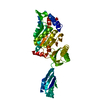
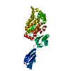
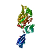
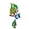
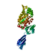
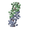
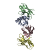
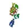
| Title | Crystal structure of Mycobacterium avium SerB2 mutant S275A/R279A at pH 6.6 with ethylene glycol bound at ACT- I domain | ||||||
 Components Components | Phosphoserine phosphatase | ||||||
 Keywords Keywords | HYDROLASE / HAD family / phosphoserine phosphatase | ||||||
| Function / homology |  Function and homology information Function and homology informationphosphoserine phosphatase / L-phosphoserine phosphatase activity / L-serine biosynthetic process / magnesium ion binding / cytoplasm Similarity search - Function | ||||||
| Biological species |  Mycobacterium avium (bacteria) Mycobacterium avium (bacteria) | ||||||
| Method |  X-RAY DIFFRACTION / SYNCHROTRON / MOLECULAR REPLACEMENT / Resolution: 3.149 Å X-RAY DIFFRACTION / SYNCHROTRON / MOLECULAR REPLACEMENT / Resolution: 3.149 Å | ||||||
 Authors Authors | Shree, S. / Dubey, S. / Ramachandran, R. | ||||||
| Funding support |  India, 1items India, 1items
| ||||||
 Citation Citation | Journal: To be published Title: Crystal Structure of Mycobacterium avium SerB2 (MAV_3907) at pH 6.6 Authors: Shree, S. / Ramachandran, R. | ||||||
| History |
|
- Structure visualization
| Structure viewer | Molecule: MolmilJmol/JSmol |
|---|
- Downloads & links
-Download
| PDBx/mmCIF format | 5t41.cif.gz | 158.3 KB | Display | PDBx/mmCIF format |
|---|---|---|---|---|
| PDB format | pdb5t41.ent.gz | 125.1 KB | Display | PDB format |
| PDBx/mmJSON format | 5t41.json.gz | Tree view | PDBx/mmJSON format | |
| Others |  Other downloads Other downloads |
-Validation report
| Arichive directory | https://data.pdbj.org/pub/pdb/validation_reports/t4/5t41ftp://data.pdbj.org/pub/pdb/validation_reports/t4/5t41 | HTTPS FTP |
|---|
-Related structure data
| Related structure data |  5is2C  5jjbC  3p96S S: Starting model for refinement C: citing same article ( |
|---|---|
| Similar structure data |
-Links
 PDBj
PDBj

- Assembly
| Deposited unit | 
| ||||||||
|---|---|---|---|---|---|---|---|---|---|
| 1 |
| ||||||||
| Unit cell |
|
-Components
| #1: Protein | Mass: 41685.703 Da / Num. of mol.: 1 / Fragment: UNP RESIDUES 5-400 / Mutation: G31R, S275A, R279A Source method: isolated from a genetically manipulated source Source: (gene. exp.) Mycobacterium avium (strain 104) (bacteria)Strain: 104 / Gene: serB, MAV_3907 / Plasmid: pAVA0421 / Production host: |
|---|---|
| #2: Chemical | ChemComp-EDO /   Mass: 62.068 Da / Num. of mol.: 1 / Source method: obtained synthetically / Formula: C2H6O2 Mass: 62.068 Da / Num. of mol.: 1 / Source method: obtained synthetically / Formula: C2H6O2 |
| #3: Chemical | ChemComp-MG /   Mass: 24.305 Da / Num. of mol.: 1 / Source method: obtained synthetically / Formula: Mg Mass: 24.305 Da / Num. of mol.: 1 / Source method: obtained synthetically / Formula: Mg |
| #4: Water | ChemComp-HOH /  Mass: 18.015 Da / Num. of mol.: 42 / Source method: isolated from a natural source / Formula: H2O Mass: 18.015 Da / Num. of mol.: 42 / Source method: isolated from a natural source / Formula: H2O |
-Experimental details
-Experiment
| Experiment | Method: X-RAY DIFFRACTION / Number of used crystals: 1 |
|---|
- Sample preparation
| Crystal | Density Matthews: 2.96 Å3/Da / Density % sol: 58.48 % Description: THE ENTRY CONTAINS FRIEDEL PAIRS IN I_PLUS/MINUS COLUMNS |
|---|---|
| Crystal grow | Temperature: 293 K / Method: vapor diffusion, hanging drop / pH: 6.6 Details: 24% PEG 8000, 0.2M Magnesium acetate tetrahydrate, 0.1M HEPES pH 6.6 |
-Data collection
| Diffraction | Mean temperature: 100 K |
|---|---|
| Diffraction source | Source: SYNCHROTRON / Site: ESRF  / Beamline: BM14 / Wavelength: 0.978 Å / Beamline: BM14 / Wavelength: 0.978 Å |
| Detector | Type: MARMOSAIC 225 mm CCD / Detector: CCD / Date: Dec 4, 2013 |
| Radiation | Protocol: SINGLE WAVELENGTH / Monochromatic (M) / Laue (L): M / Scattering type: x-ray |
| Radiation wavelength | Wavelength: 0.978 Å / Relative weight: 1 |
| Reflection | Resolution: 3.15→41.999 Å / Num. obs: 15906 / % possible obs: 95.8 % / Redundancy: 3 % / Net I/σ(I): 6.01 |
| Reflection shell | Resolution: 3.15→3.26 Å |
- Processing
| Software |
| ||||||||||||||||||||||||||||||||||||||||||||||||||||||||||||||||||||||||||||||||||||||||||||||||||||
|---|---|---|---|---|---|---|---|---|---|---|---|---|---|---|---|---|---|---|---|---|---|---|---|---|---|---|---|---|---|---|---|---|---|---|---|---|---|---|---|---|---|---|---|---|---|---|---|---|---|---|---|---|---|---|---|---|---|---|---|---|---|---|---|---|---|---|---|---|---|---|---|---|---|---|---|---|---|---|---|---|---|---|---|---|---|---|---|---|---|---|---|---|---|---|---|---|---|---|---|---|---|
| Refinement | Method to determine structure: MOLECULAR REPLACEMENT Starting model: 3P96 Resolution: 3.149→41.999 Å / SU ML: 0.42 / Cross valid method: FREE R-VALUE / σ(F): 1.34 / Phase error: 33.29 / Stereochemistry target values: ML
| ||||||||||||||||||||||||||||||||||||||||||||||||||||||||||||||||||||||||||||||||||||||||||||||||||||
| Solvent computation | Shrinkage radii: 0.9 Å / VDW probe radii: 1.11 Å / Solvent model: FLAT BULK SOLVENT MODEL | ||||||||||||||||||||||||||||||||||||||||||||||||||||||||||||||||||||||||||||||||||||||||||||||||||||
| Displacement parameters | Biso max: 89.83 Å2 / Biso mean: 62.5055 Å2 / Biso min: 27.79 Å2 | ||||||||||||||||||||||||||||||||||||||||||||||||||||||||||||||||||||||||||||||||||||||||||||||||||||
| Refinement step | Cycle: final / Resolution: 3.149→41.999 Å
| ||||||||||||||||||||||||||||||||||||||||||||||||||||||||||||||||||||||||||||||||||||||||||||||||||||
| Refine LS restraints |
| ||||||||||||||||||||||||||||||||||||||||||||||||||||||||||||||||||||||||||||||||||||||||||||||||||||
| LS refinement shell | Refine-ID: X-RAY DIFFRACTION / Total num. of bins used: 11
| ||||||||||||||||||||||||||||||||||||||||||||||||||||||||||||||||||||||||||||||||||||||||||||||||||||
| Refinement TLS params. | Method: refined / Refine-ID: X-RAY DIFFRACTION
| ||||||||||||||||||||||||||||||||||||||||||||||||||||||||||||||||||||||||||||||||||||||||||||||||||||
| Refinement TLS group |
|
