 Movie
Movie Controller

[English] 日本語
 Yorodumi
- PDB-5a04: Crystal structure of aldose-aldose oxidoreductase from Caulobacte... -
+ Open data

- Basic information
| Entry | Database: PDB / ID: 5a04 | ||||||
|---|---|---|---|---|---|---|---|
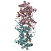
| Title | Crystal structure of aldose-aldose oxidoreductase from Caulobacter crescentus complexed with glucose | ||||||
 Components Components | ALDOSE-ALDOSE OXIDOREDUCTASE | ||||||
 Keywords Keywords | OXIDOREDUCTASE | ||||||
| Function / homology |  Function and homology information Function and homology information | ||||||
| Biological species |  CAULOBACTER CRESCENTUS CB15 (bacteria) CAULOBACTER CRESCENTUS CB15 (bacteria) | ||||||
| Method |  X-RAY DIFFRACTION / SYNCHROTRON / MOLECULAR REPLACEMENT / Resolution: 1.698 Å X-RAY DIFFRACTION / SYNCHROTRON / MOLECULAR REPLACEMENT / Resolution: 1.698 Å | ||||||
 Authors Authors | Taberman, H. / Rouvinen, J. / Parkkinen, T. | ||||||
 Citation Citation | Journal: Biochem.J. / Year: 2015 Title: Structure and Function of Caulobacter Crescentus Aldose-Aldose Oxidoreductase. Authors: Taberman, H. / Andberg, M. / Koivula, A. / Hakulinen, N. / Penttila, M. / Rouvinen, J. / Parkkinen, T. | ||||||
| History |
|
- Structure visualization
| Structure viewer | Molecule: MolmilJmol/JSmol |
|---|
- Downloads & links
-Download
| PDBx/mmCIF format | 5a04.cif.gz | 438.1 KB | Display | PDBx/mmCIF format |
|---|---|---|---|---|
| PDB format | pdb5a04.ent.gz | 361.7 KB | Display | PDB format |
| PDBx/mmJSON format | 5a04.json.gz | Tree view | PDBx/mmJSON format | |
| Others |  Other downloads Other downloads |
-Validation report
| Arichive directory | https://data.pdbj.org/pub/pdb/validation_reports/a0/5a04ftp://data.pdbj.org/pub/pdb/validation_reports/a0/5a04 | HTTPS FTP |
|---|
-Related structure data
| Related structure data |  5a02C  5a03C  5a05C  5a06C  1h6aS C: citing same article ( S: Starting model for refinement |
|---|---|
| Similar structure data |


-Links
 PDBj
PDBj


- Assembly
| Deposited unit | 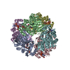
| ||||||||||||||||||||||||
|---|---|---|---|---|---|---|---|---|---|---|---|---|---|---|---|---|---|---|---|---|---|---|---|---|---|
| 1 | 
| ||||||||||||||||||||||||
| 2 | 
| ||||||||||||||||||||||||
| 3 | 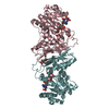
| ||||||||||||||||||||||||
| Unit cell |
| ||||||||||||||||||||||||
| Noncrystallographic symmetry (NCS) | NCS oper:
|
-Components
-Protein / Sugars , 2 types, 15 molecules ABCDEF
| #1: Protein | Mass: 37289.438 Da / Num. of mol.: 6 Source method: isolated from a genetically manipulated source Source: (gene. exp.) CAULOBACTER CRESCENTUS CB15 (bacteria) / Strain: CB15 / Production host:  References: UniProt: Q9A8X3*PLUS, Oxidoreductases; Acting on the CH-OH group of donors; With unknown physiological acceptors #3: Sugar | ChemComp-BGC /  Type: D-saccharide, beta linking / Mass: 180.156 Da / Num. of mol.: 9 Type: D-saccharide, beta linking / Mass: 180.156 Da / Num. of mol.: 9Source method: isolated from a genetically manipulated source Formula: C6H12O6 |
|---|
-Non-polymers , 4 types, 2054 molecules 


| #2: Chemical | ChemComp-NDP /  Mass: 745.421 Da / Num. of mol.: 6 / Source method: obtained synthetically / Formula: C21H30N7O17P3 Mass: 745.421 Da / Num. of mol.: 6 / Source method: obtained synthetically / Formula: C21H30N7O17P3#4: Chemical | ChemComp-DIO /  Mass: 88.105 Da / Num. of mol.: 5 / Source method: obtained synthetically / Formula: C4H8O2 Mass: 88.105 Da / Num. of mol.: 5 / Source method: obtained synthetically / Formula: C4H8O2#5: Chemical | ChemComp-SO4 /  Mass: 96.063 Da / Num. of mol.: 6 / Source method: obtained synthetically / Formula: SO4 Mass: 96.063 Da / Num. of mol.: 6 / Source method: obtained synthetically / Formula: SO4#6: Water | ChemComp-HOH / | Mass: 18.015 Da / Num. of mol.: 2037 / Source method: isolated from a natural source / Formula: H2O |
|---|
-Experimental details
-Experiment
| Experiment | Method: X-RAY DIFFRACTION / Number of used crystals: 1 |
|---|
- Sample preparation
| Crystal | Density Matthews: 3.6 Å3/Da / Density % sol: 66 % / Description: NONE |
|---|---|
| Crystal grow | pH: 6.5 / Details: MES PH 6.5, AMMONIUM SULPHATE, DIOXANE |
-Data collection
| Diffraction | Mean temperature: 100 K |
|---|---|
| Diffraction source | Source: SYNCHROTRON / Site: ESRF  / Beamline: ID29 / Wavelength: 0.97625 / Beamline: ID29 / Wavelength: 0.97625 |
| Detector | Type: DECTRIS PILATUS 6M / Detector: PIXEL / Date: Feb 27, 2014 / Details: MIRRORS |
| Radiation | Monochromator: CHANNEL-CUT SI / Protocol: SINGLE WAVELENGTH / Monochromatic (M) / Laue (L): M / Scattering type: x-ray |
| Radiation wavelength | Wavelength: 0.97625 Å / Relative weight: 1 |
| Reflection | Resolution: 1.7→50 Å / Num. obs: 338561 / % possible obs: 99 % / Observed criterion σ(I): 2 / Redundancy: 3.5 % / Biso Wilson estimate: 21.3 Å2 / Rmerge(I) obs: 0.06 / Net I/σ(I): 13 |
| Reflection shell | Resolution: 1.7→1.8 Å / Redundancy: 3.5 % / Rmerge(I) obs: 0.6 / Mean I/σ(I) obs: 2 / % possible all: 97 |
- Processing
| Software |
| |||||||||||||||||||||||||||||||||||||||||||||||||||||||||||||||||||||||||||||||||||||||||||||||||||||||||||||||||||||||||||||||||||||||||||||||||||||||||||||||||||||||||||||||||||||||||||||||||||||||||||||||||||||||||
|---|---|---|---|---|---|---|---|---|---|---|---|---|---|---|---|---|---|---|---|---|---|---|---|---|---|---|---|---|---|---|---|---|---|---|---|---|---|---|---|---|---|---|---|---|---|---|---|---|---|---|---|---|---|---|---|---|---|---|---|---|---|---|---|---|---|---|---|---|---|---|---|---|---|---|---|---|---|---|---|---|---|---|---|---|---|---|---|---|---|---|---|---|---|---|---|---|---|---|---|---|---|---|---|---|---|---|---|---|---|---|---|---|---|---|---|---|---|---|---|---|---|---|---|---|---|---|---|---|---|---|---|---|---|---|---|---|---|---|---|---|---|---|---|---|---|---|---|---|---|---|---|---|---|---|---|---|---|---|---|---|---|---|---|---|---|---|---|---|---|---|---|---|---|---|---|---|---|---|---|---|---|---|---|---|---|---|---|---|---|---|---|---|---|---|---|---|---|---|---|---|---|---|---|---|---|---|---|---|---|---|---|---|---|---|---|---|---|---|
| Refinement | Method to determine structure: MOLECULAR REPLACEMENT Starting model: PDB ENTRY 1H6A Resolution: 1.698→47.591 Å / SU ML: 0.2 / σ(F): 1.36 / Phase error: 17.9 / Stereochemistry target values: ML
| |||||||||||||||||||||||||||||||||||||||||||||||||||||||||||||||||||||||||||||||||||||||||||||||||||||||||||||||||||||||||||||||||||||||||||||||||||||||||||||||||||||||||||||||||||||||||||||||||||||||||||||||||||||||||
| Solvent computation | Shrinkage radii: 0.9 Å / VDW probe radii: 1.11 Å / Solvent model: FLAT BULK SOLVENT MODEL | |||||||||||||||||||||||||||||||||||||||||||||||||||||||||||||||||||||||||||||||||||||||||||||||||||||||||||||||||||||||||||||||||||||||||||||||||||||||||||||||||||||||||||||||||||||||||||||||||||||||||||||||||||||||||
| Displacement parameters | Biso mean: 24.2 Å2 | |||||||||||||||||||||||||||||||||||||||||||||||||||||||||||||||||||||||||||||||||||||||||||||||||||||||||||||||||||||||||||||||||||||||||||||||||||||||||||||||||||||||||||||||||||||||||||||||||||||||||||||||||||||||||
| Refinement step | Cycle: LAST / Resolution: 1.698→47.591 Å
| |||||||||||||||||||||||||||||||||||||||||||||||||||||||||||||||||||||||||||||||||||||||||||||||||||||||||||||||||||||||||||||||||||||||||||||||||||||||||||||||||||||||||||||||||||||||||||||||||||||||||||||||||||||||||
| Refine LS restraints |
| |||||||||||||||||||||||||||||||||||||||||||||||||||||||||||||||||||||||||||||||||||||||||||||||||||||||||||||||||||||||||||||||||||||||||||||||||||||||||||||||||||||||||||||||||||||||||||||||||||||||||||||||||||||||||
| LS refinement shell |
|
