+ Open data
- Basic information
| Entry | Database: PDB / ID: 5irz | |||||||||||||||
|---|---|---|---|---|---|---|---|---|---|---|---|---|---|---|---|---|
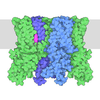
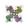
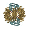
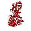
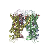
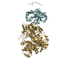
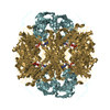
| Title | Structure of TRPV1 determined in lipid nanodisc | |||||||||||||||
 Components Components | Transient receptor potential cation channel subfamily V member 1 | |||||||||||||||
 Keywords Keywords | TRANSPORT PROTEIN / TRP / ion channel / nanodisc / vanilloid / lipid | |||||||||||||||
| Function / homology |  Function and homology information Function and homology informationnegative regulation of establishment of blood-brain barrier / response to capsazepine / sensory perception of mechanical stimulus / cellular response to temperature stimulus / peptide secretion / smooth muscle contraction involved in micturition / excitatory extracellular ligand-gated monoatomic ion channel activity / temperature-gated ion channel activity / detection of chemical stimulus involved in sensory perception of pain / TRP channels ...negative regulation of establishment of blood-brain barrier / response to capsazepine / sensory perception of mechanical stimulus / cellular response to temperature stimulus / peptide secretion / smooth muscle contraction involved in micturition / excitatory extracellular ligand-gated monoatomic ion channel activity / temperature-gated ion channel activity / detection of chemical stimulus involved in sensory perception of pain / TRP channels / fever generation / detection of temperature stimulus involved in thermoception / urinary bladder smooth muscle contraction / thermoception / cellular response to acidic pH / negative regulation of systemic arterial blood pressure / response to pH / chloride channel regulator activity / dendritic spine membrane / monoatomic cation transmembrane transporter activity / glutamate secretion / ligand-gated monoatomic ion channel activity / negative regulation of heart rate / response to pain / temperature homeostasis / diet induced thermogenesis / cellular response to alkaloid / cellular response to ATP / cellular response to cytokine stimulus / detection of temperature stimulus involved in sensory perception of pain / intracellularly gated calcium channel activity / behavioral response to pain / negative regulation of mitochondrial membrane potential / calcium ion import across plasma membrane / monoatomic cation channel activity / extracellular ligand-gated monoatomic ion channel activity / sensory perception of pain / phosphatidylinositol binding / phosphoprotein binding / microglial cell activation / cellular response to nerve growth factor stimulus / response to peptide hormone / calcium ion transmembrane transport / GABA-ergic synapse / lipid metabolic process / cellular response to growth factor stimulus / calcium channel activity / positive regulation of nitric oxide biosynthetic process / cellular response to tumor necrosis factor / transmembrane signaling receptor activity / calcium ion transport / sensory perception of taste / cellular response to heat / positive regulation of cytosolic calcium ion concentration / response to heat / monoatomic ion transmembrane transport / protein homotetramerization / postsynaptic membrane / calmodulin binding / neuron projection / positive regulation of apoptotic process / external side of plasma membrane / neuronal cell body / dendrite / negative regulation of transcription by RNA polymerase II / ATP binding / metal ion binding / identical protein binding / membrane / plasma membrane Similarity search - Function | |||||||||||||||
| Biological species |  | |||||||||||||||
| Method | ELECTRON MICROSCOPY / single particle reconstruction / cryo EM / Resolution: 3.28 Å | |||||||||||||||
 Authors Authors | Gao, Y. / Cao, E. / Julius, D. / Cheng, Y. | |||||||||||||||
| Funding support |  United States, 4items United States, 4items
| |||||||||||||||
 Citation Citation |  Journal: Nature / Year: 2016 Journal: Nature / Year: 2016Title: TRPV1 structures in nanodiscs reveal mechanisms of ligand and lipid action. Authors: Yuan Gao / Erhu Cao / David Julius / Yifan Cheng /  Abstract: When integral membrane proteins are visualized in detergents or other artificial systems, an important layer of information is lost regarding lipid interactions and their effects on protein structure. ...When integral membrane proteins are visualized in detergents or other artificial systems, an important layer of information is lost regarding lipid interactions and their effects on protein structure. This is especially relevant to proteins for which lipids have both structural and regulatory roles. Here we demonstrate the power of combining electron cryo-microscopy with lipid nanodisc technology to ascertain the structure of the rat TRPV1 ion channel in a native bilayer environment. Using this approach, we determined the locations of annular and regulatory lipids and showed that specific phospholipid interactions enhance binding of a spider toxin to TRPV1 through formation of a tripartite complex. Furthermore, phosphatidylinositol lipids occupy the binding site for capsaicin and other vanilloid ligands, suggesting a mechanism whereby chemical or thermal stimuli elicit channel activation by promoting the release of bioactive lipids from a critical allosteric regulatory site. | |||||||||||||||
| History |
|
- Structure visualization
| Movie |
 Movie viewer Movie viewer |
|---|---|
| Structure viewer | Molecule:  Molmil Molmil Jmol/JSmol Jmol/JSmol |
- Downloads & links
- Download
| PDBx/mmCIF format |  5irz.cif.gz 5irz.cif.gz | 319.4 KB | Display |  PDBx/mmCIF format PDBx/mmCIF format |
|---|---|---|---|---|
| PDB format |  pdb5irz.ent.gz pdb5irz.ent.gz | 246.3 KB | Display |  PDB format PDB format |
| PDBx/mmJSON format |  5irz.json.gz 5irz.json.gz | Tree view |  PDBx/mmJSON format PDBx/mmJSON format | |
| Others |  Other downloads Other downloads |
-Validation report
| Arichive directory |  https://data.pdbj.org/pub/pdb/validation_reports/ir/5irz https://data.pdbj.org/pub/pdb/validation_reports/ir/5irz ftp://data.pdbj.org/pub/pdb/validation_reports/ir/5irz ftp://data.pdbj.org/pub/pdb/validation_reports/ir/5irz | HTTPS FTP |
|---|
-Related structure data
| Related structure data |  8118MC  8117C  8119C  8120C  5irxC  5is0C M: map data used to model this data C: citing same article ( |
|---|---|
| Similar structure data |
- Links
Links
- Assembly
| Deposited unit | 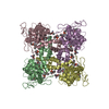
|
|---|---|
| 1 |
|
- Components
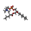
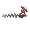
| #1: Protein | Mass: 72959.297 Da / Num. of mol.: 4 / Fragment: UNP residues 110-603, 627-764 Source method: isolated from a genetically manipulated source Source: (gene. exp.)   Homo sapiens (human) / References: UniProt: O35433 Homo sapiens (human) / References: UniProt: O35433#2: Chemical | ChemComp-6O8 / ( #3: Chemical | ChemComp-6ES / ( #4: Chemical | ChemComp-6OE / ( |
|---|
-Experimental details
-Experiment
| Experiment | Method: ELECTRON MICROSCOPY |
|---|---|
| EM experiment | Aggregation state: PARTICLE / 3D reconstruction method: single particle reconstruction |
- Sample preparation
| Component | Name: TRPV1 ion channel in unliganded state / Type: COMPLEX / Entity ID: #1 / Source: RECOMBINANT | ||||||||||||||||||||
|---|---|---|---|---|---|---|---|---|---|---|---|---|---|---|---|---|---|---|---|---|---|
| Source (natural) | Organism:  | ||||||||||||||||||||
| Source (recombinant) | Organism:  Homo sapiens (human) / Plasmid: unknown Homo sapiens (human) / Plasmid: unknown | ||||||||||||||||||||
| Buffer solution | pH: 7.4 | ||||||||||||||||||||
| Buffer component |
| ||||||||||||||||||||
| Specimen | Conc.: 0.5 mg/ml / Embedding applied: NO / Shadowing applied: NO / Staining applied: NO / Vitrification applied: YES / Details: This sample was monodisperse. | ||||||||||||||||||||
| Specimen support | Grid material: COPPER / Grid mesh size: 400 divisions/in. / Grid type: Quantifoil R1.2/1.3 | ||||||||||||||||||||
| Vitrification | Instrument: FEI VITROBOT MARK III / Cryogen name: ETHANE / Humidity: 100 % / Chamber temperature: 278 K Details: Blot for 6 seconds before plunging into liquid ethane (FEI VITROBOT MARK III). |
- Electron microscopy imaging
| Experimental equipment |  Model: Tecnai Polara / Image courtesy: FEI Company |
|---|---|
| Microscopy | Model: FEI POLARA 300 / Details: Grid screening was performed manually. |
| Electron gun | Electron source:  FIELD EMISSION GUN / Accelerating voltage: 300 kV / Illumination mode: FLOOD BEAM FIELD EMISSION GUN / Accelerating voltage: 300 kV / Illumination mode: FLOOD BEAM |
| Electron lens | Mode: BRIGHT FIELD / Nominal magnification: 31000 X / Calibrated magnification: 41132 X / Nominal defocus max: 2200 nm / Nominal defocus min: 700 nm / Calibrated defocus min: 700 nm / Calibrated defocus max: 2200 nm / Cs: 2 mm / C2 aperture diameter: 30 µm / Alignment procedure: COMA FREE |
| Specimen holder | Cryogen: NITROGEN / Specimen holder model: OTHER / Temperature (max): 70 K / Temperature (min): 70 K |
| Image recording | Average exposure time: 6 sec. / Electron dose: 41 e/Å2 / Detector mode: SUPER-RESOLUTION / Film or detector model: GATAN K2 SUMMIT (4k x 4k) / Num. of grids imaged: 1 / Num. of real images: 1000 Details: Images were collected in movie mode at 5 frames per second for 6 seconds. |
| Image scans | Width: 3838 / Height: 3710 / Movie frames/image: 30 / Used frames/image: 1-30 |
- Processing
| Software | Name: PHENIX / Version: 1.10_2152: / Classification: refinement | |||||||||||||||||||||||||||||||||||||||||||||||||||||||
|---|---|---|---|---|---|---|---|---|---|---|---|---|---|---|---|---|---|---|---|---|---|---|---|---|---|---|---|---|---|---|---|---|---|---|---|---|---|---|---|---|---|---|---|---|---|---|---|---|---|---|---|---|---|---|---|---|
| EM software |
| |||||||||||||||||||||||||||||||||||||||||||||||||||||||
| Image processing | Details: Each image stack was subjected to whole-frame motion correction followed by correction at the individual pixel level using program UcsfDfCorr. | |||||||||||||||||||||||||||||||||||||||||||||||||||||||
| CTF correction | Details: CTF correction was performed before classification and refinement. Type: PHASE FLIPPING AND AMPLITUDE CORRECTION | |||||||||||||||||||||||||||||||||||||||||||||||||||||||
| Particle selection | Num. of particles selected: 159193 Details: Initial manual particle picking and automatic particle picking were performed using SamViewer and several python scripts based on SPIDER. | |||||||||||||||||||||||||||||||||||||||||||||||||||||||
| Symmetry | Point symmetry: C4 (4 fold cyclic) | |||||||||||||||||||||||||||||||||||||||||||||||||||||||
| 3D reconstruction | Resolution: 3.28 Å / Resolution method: FSC 0.143 CUT-OFF / Num. of particles: 30689 / Num. of class averages: 1 / Symmetry type: POINT | |||||||||||||||||||||||||||||||||||||||||||||||||||||||
| Atomic model building | Protocol: RIGID BODY FIT / Space: REAL / Target criteria: correlation coefficient | |||||||||||||||||||||||||||||||||||||||||||||||||||||||
| Atomic model building | PDB-ID: 3J5P Pdb chain-ID: A / Accession code: 3J5P / Source name: PDB / Type: experimental model | |||||||||||||||||||||||||||||||||||||||||||||||||||||||
| Refinement | Highest resolution: 3.28 Å | |||||||||||||||||||||||||||||||||||||||||||||||||||||||
| Refine LS restraints |
|
 Movie
Movie Controller




 PDBj
PDBj