 Movie
Movie Controller

[English] 日本語
 Yorodumi
- PDB-3j5p: Structure of TRPV1 ion channel determined by single particle elec... -
+ Open data

- Basic information
| Entry | Database: PDB / ID: 3j5p | ||||||
|---|---|---|---|---|---|---|---|
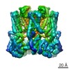
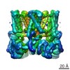
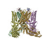
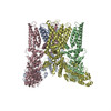
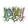
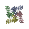
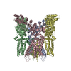
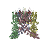
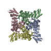
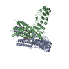
| Title | Structure of TRPV1 ion channel determined by single particle electron cryo-microscopy | ||||||
 Components Components | Transient receptor potential cation channel subfamily V member 1 | ||||||
 Keywords Keywords | TRANSPORT PROTEIN / TRPV1 channel | ||||||
| Function / homology |  Function and homology information Function and homology informationnegative regulation of iodide transmembrane transport / positive regulation of membrane depolarization / negative regulation of establishment of blood-brain barrier / response to capsazepine / sensory perception of mechanical stimulus / cellular response to temperature stimulus / peptide secretion / positive regulation of sensory perception of pain / temperature-gated ion channel activity / detection of chemical stimulus involved in sensory perception of pain ...negative regulation of iodide transmembrane transport / positive regulation of membrane depolarization / negative regulation of establishment of blood-brain barrier / response to capsazepine / sensory perception of mechanical stimulus / cellular response to temperature stimulus / peptide secretion / positive regulation of sensory perception of pain / temperature-gated ion channel activity / detection of chemical stimulus involved in sensory perception of pain / positive regulation of renal sodium excretion / TRP channels / negative regulation of axon regeneration / positive regulation of cardiac muscle cell differentiation / smooth muscle contraction involved in micturition / fever generation / detection of temperature stimulus involved in thermoception / urinary bladder smooth muscle contraction / thermoception / response to pH / glutamate secretion / monoatomic cation transmembrane transporter activity / negative regulation of systemic arterial blood pressure / excitatory extracellular ligand-gated monoatomic ion channel activity / dendritic spine membrane / negative regulation of heart rate / response to acidic pH / positive regulation of urine volume / response to pain / diet induced thermogenesis / cellular response to alkaloid / cellular response to cytokine stimulus / temperature homeostasis / intracellularly gated calcium channel activity / cellular response to ATP / negative regulation of mitochondrial membrane potential / detection of temperature stimulus involved in sensory perception of pain / calcium ion import across plasma membrane / behavioral response to pain / positive regulation of vasoconstriction / monoatomic ion channel activity / ligand-gated monoatomic ion channel activity / monoatomic cation channel activity / cellular response to acidic pH / extracellular ligand-gated monoatomic ion channel activity / phosphatidylinositol binding / sensory perception of pain / axon terminus / positive regulation of excitatory postsynaptic potential / sarcoplasmic reticulum / lipid metabolic process / phosphoprotein binding / microglial cell activation / cellular response to nerve growth factor stimulus / cellular response to growth factor stimulus / cellular response to tumor necrosis factor / response to peptide hormone / GABA-ergic synapse / calcium ion transmembrane transport / calcium channel activity / positive regulation of nitric oxide biosynthetic process / calcium ion transport / transmembrane signaling receptor activity / cellular response to heat / sensory perception of taste / response to heat / positive regulation of cytosolic calcium ion concentration / monoatomic ion transmembrane transport / protein homotetramerization / calmodulin binding / postsynaptic membrane / neuron projection / positive regulation of apoptotic process / external side of plasma membrane / neuronal cell body / dendrite / negative regulation of transcription by RNA polymerase II / ATP binding / membrane / metal ion binding / identical protein binding / nucleus / plasma membrane Similarity search - Function | ||||||
| Biological species |  | ||||||
| Method | ELECTRON MICROSCOPY / single particle reconstruction / cryo EM / Resolution: 3.275 Å | ||||||
 Authors Authors | Liao, M. / Cao, E. / Julius, D. / Cheng, Y. | ||||||
 Citation Citation | Journal: Nature / Year: 2013 Title: Structure of the TRPV1 ion channel determined by electron cryo-microscopy. Authors: Maofu Liao / Erhu Cao / David Julius / Yifan Cheng /  Abstract: Transient receptor potential (TRP) channels are sensors for a wide range of cellular and environmental signals, but elucidating how these channels respond to physical and chemical stimuli has been ...Transient receptor potential (TRP) channels are sensors for a wide range of cellular and environmental signals, but elucidating how these channels respond to physical and chemical stimuli has been hampered by a lack of detailed structural information. Here we exploit advances in electron cryo-microscopy to determine the structure of a mammalian TRP channel, TRPV1, at 3.4 Å resolution, breaking the side-chain resolution barrier for membrane proteins without crystallization. Like voltage-gated channels, TRPV1 exhibits four-fold symmetry around a central ion pathway formed by transmembrane segments 5-6 (S5-S6) and the intervening pore loop, which is flanked by S1-S4 voltage-sensor-like domains. TRPV1 has a wide extracellular 'mouth' with a short selectivity filter. The conserved 'TRP domain' interacts with the S4-S5 linker, consistent with its contribution to allosteric modulation. Subunit organization is facilitated by interactions among cytoplasmic domains, including amino-terminal ankyrin repeats. These observations provide a structural blueprint for understanding unique aspects of TRP channel function. | ||||||
| History |
|
- Structure visualization
| Movie |
Movie viewer |
|---|---|
| Structure viewer | Molecule: MolmilJmol/JSmol |
- Downloads & links
-Download
| PDBx/mmCIF format | 3j5p.cif.gz | 416.4 KB | Display | PDBx/mmCIF format |
|---|---|---|---|---|
| PDB format | pdb3j5p.ent.gz | 337.5 KB | Display | PDB format |
| PDBx/mmJSON format | 3j5p.json.gz | Tree view | PDBx/mmJSON format | |
| Others |  Other downloads Other downloads |
-Validation report
| Arichive directory | https://data.pdbj.org/pub/pdb/validation_reports/j5/3j5pftp://data.pdbj.org/pub/pdb/validation_reports/j5/3j5p | HTTPS FTP |
|---|
-Related structure data
| Related structure data | 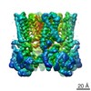 5778MC M: map data used to model this data C: citing same article ( |
|---|---|
| Similar structure data | |
| EM raw data | EMPIAR-10005 (Title: TRPV1 dataset taken on a K2 direct electron detector / Data size: 6.3 TB / Data #1: TRPV1 picked particles [tilt series] Data #2: TRPV1 raw multi-frame micrographs [micrographs - multiframe] Data #3: TRPV1 summed frame micrographs [micrographs - single frame]) |


-Links
 PDBj
PDBj

- Assembly
| Deposited unit | 
|
|---|---|
| 1 |
|
-Components
| #1: Protein | Mass: 68242.156 Da / Num. of mol.: 4 / Fragment: SEE REMARK 999 Source method: isolated from a genetically manipulated source Source: (gene. exp.)  Homo sapiens (human) / References: UniProt: O35433 Homo sapiens (human) / References: UniProt: O35433Sequence details | THE TRPV1 CONSTRUCT COMPRISES RESIDUES 110-603 AND 627-764, WITH RESIDUES 604-626 DELETED. RESIDUES ...THE TRPV1 CONSTRUCT COMPRISES RESIDUES 110-603 AND 627-764, WITH RESIDUES 604-626 DELETED. RESIDUES 719-764 ARE NOT MODELED, WITH THE EXCEPTION OF 11 RESIDUES (NUMBERED 752-762 IN THE COORDINATE | |
|---|
-Experimental details
-Experiment
| Experiment | Method: ELECTRON MICROSCOPY |
|---|---|
| EM experiment | Aggregation state: PARTICLE / 3D reconstruction method: single particle reconstruction |
- Sample preparation
| Component | Name: Rat TRPV1 / Type: COMPLEX / Details: Tetramer |
|---|---|
| Molecular weight | Value: 0.3 MDa / Experimental value: NO |
| Buffer solution | Name: 150 mM NaCl, 20 mM HEPES, 2 mM TCEP / pH: 7.4 / Details: 150 mM NaCl, 20 mM HEPES, 2 mM TCEP |
| Specimen | Conc.: 0.3 mg/ml / Embedding applied: NO / Shadowing applied: NO / Staining applied: NO / Vitrification applied: YES |
| Specimen support | Details: 400 mesh Quantifoil grid |
| Vitrification | Instrument: FEI VITROBOT MARK III / Cryogen name: ETHANE / Temp: 120 K / Humidity: 90 % Details: Blot for 6 seconds before plunging into liquid ethane (FEI VITROBOT MARK III) Method: Blot for 6 sec |
- Electron microscopy imaging
| Experimental equipment |  Model: Tecnai Polara / Image courtesy: FEI Company |
|---|---|
| Microscopy | Model: FEI POLARA 300 / Date: Jan 1, 2013 Details: K2 Summit in super-resolution counting mode. Motion correction as described in Li et al. (2013) Nature Methods. |
| Electron gun | Electron source:  FIELD EMISSION GUN / Accelerating voltage: 300 kV / Illumination mode: FLOOD BEAM FIELD EMISSION GUN / Accelerating voltage: 300 kV / Illumination mode: FLOOD BEAM |
| Electron lens | Mode: BRIGHT FIELD / Nominal magnification: 31000 X / Calibrated magnification: 31000 X / Nominal defocus max: 3000 nm / Nominal defocus min: 1500 nm / Cs: 2 mm / Camera length: 0 mm |
| Specimen holder | Specimen holder model: OTHER / Specimen holder type: FEI Polara cartridge |
| Image recording | Electron dose: 21 e/Å2 / Film or detector model: GATAN K2 SUMMIT (4k x 4k) Details: Operated in super-resolution counting mode, dose fractionation |
| Image scans | Num. digital images: 946 |
| Radiation | Protocol: SINGLE WAVELENGTH / Monochromatic (M) / Laue (L): M / Scattering type: x-ray |
| Radiation wavelength | Relative weight: 1 |
- Processing
| EM software | Name: RELION / Category: 3D reconstruction | ||||||||||||
|---|---|---|---|---|---|---|---|---|---|---|---|---|---|
| CTF correction | Details: Each particle | ||||||||||||
| Symmetry | Point symmetry: C4 (4 fold cyclic) | ||||||||||||
| 3D reconstruction | Method: Maximum likelihood / Resolution: 3.275 Å / Resolution method: FSC 0.143 CUT-OFF / Num. of particles: 35645 / Nominal pixel size: 1.2156 Å / Actual pixel size: 1.2156 Å Details: The entire ankyrin structure was fitted as a single rigid body. As a result, the first two ankyrin repeats (UNP residues 111-199) are included but are not well defined by the experimental ...Details: The entire ankyrin structure was fitted as a single rigid body. As a result, the first two ankyrin repeats (UNP residues 111-199) are included but are not well defined by the experimental density. (Single particle details: 3D classification, refinement, and reconstruction were performed using RELION) (Single particle--Applied symmetry: C4) Refinement type: HALF-MAPS REFINED INDEPENDENTLY / Symmetry type: POINT | ||||||||||||
| Atomic model building | Protocol: AB INITIO MODEL / Space: REAL Details: REFINEMENT PROTOCOL--de novo model building DETAILS--The entire ankyrin structure was fitted as a single rigid body. As a result, the first two ankyrin repeats (UNP residues 111-199) are ...Details: REFINEMENT PROTOCOL--de novo model building DETAILS--The entire ankyrin structure was fitted as a single rigid body. As a result, the first two ankyrin repeats (UNP residues 111-199) are included but are not well defined by the experimental density. | ||||||||||||
| Refinement step | Cycle: LAST
|
