[English] 日本語
Yorodumi
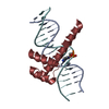
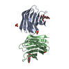
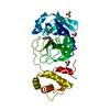
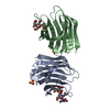
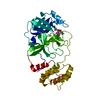
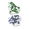
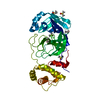
- PDB-5hod: Structure of LHX4 transcription factor complexed with DNA -

+
Open data


ID or keywords:

Loading...

-
Basic information

Entry
Database: PDB / ID: 5hod
TitleStructure of LHX4 transcription factor complexed with DNA
Components
  • DNA (5'-D(P*AP*CP*CP*TP*AP*AP*TP*TP*AP*GP*GP*CP*GP*TP*AP*AP*TP*TP*AP*G)-3')
  • DNA (5'-D(P*CP*TP*AP*AP*TP*TP*AP*CP*GP*CP*CP*TP*AP*AP*TP*TP*AP*GP*GP*T)-3')
  • LIM/homeobox protein Lhx4
KeywordsTRANSCRIPTION / Transcription factor / Complex with DNA / Dimer
Function / homology
Function and homology information


medial motor column neuron differentiation / methyl-CpG binding / motor neuron axon guidance / placenta development / RNA polymerase II transcription regulatory region sequence-specific DNA binding / animal organ morphogenesis / neuron differentiation / Regulation of expression of SLITs and ROBOs / sequence-specific double-stranded DNA binding / DNA-binding transcription activator activity, RNA polymerase II-specific ...medial motor column neuron differentiation / methyl-CpG binding / motor neuron axon guidance / placenta development / RNA polymerase II transcription regulatory region sequence-specific DNA binding / animal organ morphogenesis / neuron differentiation / Regulation of expression of SLITs and ROBOs / sequence-specific double-stranded DNA binding / DNA-binding transcription activator activity, RNA polymerase II-specific / sequence-specific DNA binding / DNA-binding transcription factor activity, RNA polymerase II-specific / chromatin / regulation of transcription by RNA polymerase II / negative regulation of apoptotic process / positive regulation of transcription by RNA polymerase II / metal ion binding / nucleus
Similarity search - Function
LIM zinc-binding domain signature. / LIM domain / Zinc-binding domain present in Lin-11, Isl-1, Mec-3. / Zinc finger, LIM-type / LIM domain profile. / Homeobox, conserved site / 'Homeobox' domain signature. / Homeodomain / 'Homeobox' domain profile. / Homeodomain ...LIM zinc-binding domain signature. / LIM domain / Zinc-binding domain present in Lin-11, Isl-1, Mec-3. / Zinc finger, LIM-type / LIM domain profile. / Homeobox, conserved site / 'Homeobox' domain signature. / Homeodomain / 'Homeobox' domain profile. / Homeodomain / Homeobox domain / Homeodomain-like / Homeobox-like domain superfamily / Arc Repressor Mutant, subunit A / Orthogonal Bundle / Mainly Alpha
Similarity search - Domain/homology
DNA / DNA (> 10) / LIM/homeobox protein Lhx4
Similarity search - Component
Biological speciesHomo sapiens (human)
MethodX-RAY DIFFRACTION / SYNCHROTRON / MOLECULAR REPLACEMENT / Resolution: 2.682 Å
AuthorsMorgunova, E. / Yin, Y. / Popov, A. / Taipale, J.
CitationJournal: Science / Year: 2017
Title: Impact of cytosine methylation on DNA binding specificities of human transcription factors.
Authors: Yin, Y. / Morgunova, E. / Jolma, A. / Kaasinen, E. / Sahu, B. / Khund-Sayeed, S. / Das, P.K. / Kivioja, T. / Dave, K. / Zhong, F. / Nitta, K.R. / Taipale, M. / Popov, A. / Ginno, P.A. / ...Authors: Yin, Y. / Morgunova, E. / Jolma, A. / Kaasinen, E. / Sahu, B. / Khund-Sayeed, S. / Das, P.K. / Kivioja, T. / Dave, K. / Zhong, F. / Nitta, K.R. / Taipale, M. / Popov, A. / Ginno, P.A. / Domcke, S. / Yan, J. / Schubeler, D. / Vinson, C. / Taipale, J.
History
DepositionJan 19, 2016Deposition site: RCSB / Processing site: PDBE
Revision 1.0Feb 1, 2017Provider: repository / Type: Initial release
Revision 1.1May 17, 2017Group: Database references
Revision 1.2Jan 10, 2024Group: Data collection / Database references / Refinement description
Category: chem_comp_atom / chem_comp_bond ...chem_comp_atom / chem_comp_bond / database_2 / pdbx_initial_refinement_model
Item: _database_2.pdbx_DOI / _database_2.pdbx_database_accession

-
Structure visualization

Structure viewerMolecule:
MolmilJmol/JSmol

Downloads & links

-
Assembly

Deposited unit
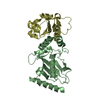
A: LIM/homeobox protein Lhx4
B: DNA (5'-D(P*AP*CP*CP*TP*AP*AP*TP*TP*AP*GP*GP*CP*GP*TP*AP*AP*TP*TP*AP*G)-3')
C: DNA (5'-D(P*CP*TP*AP*AP*TP*TP*AP*CP*GP*CP*CP*TP*AP*AP*TP*TP*AP*GP*GP*T)-3')
D: LIM/homeobox protein Lhx4


Theoretical massNumber of molelcules
Total (without water)26,5944
Polymers26,5944
Non-polymers00
Water48627
1


  • Idetical with deposited unit
  • defined by author&software
TypeNameSymmetry operationNumber
identity operation1_555x,y,z1
Buried area5250 Å2
ΔGint-23 kcal/mol
Surface area14140 Å2
MethodPISA
Unit cell
Length a, b, c (Å)40.768, 68.442, 200.464
Angle α, β, γ (deg.)90.00, 90.00, 90.00
Int Tables number20
Space group name H-MC2221
Components on special symmetry positions
IDModelComponents
11A-215-

HOH

-
Components

#1: Protein LIM/homeobox protein Lhx4 / LIM homeobox protein 4


Mass: 7164.329 Da / Num. of mol.: 2 / Fragment: UNP residues 156-216
Source method: isolated from a genetically manipulated source
Source: (gene. exp.) Homo sapiens (human) / Gene: LHX4 / Plasmid: pETG20A / Production host: Escherichia coli BL21(DE3) (bacteria) / Variant (production host): Rosetta / References: UniProt: Q969G2
#2: DNA chain DNA (5'-D(P*AP*CP*CP*TP*AP*AP*TP*TP*AP*GP*GP*CP*GP*TP*AP*AP*TP*TP*AP*G)-3')


Mass: 6157.017 Da / Num. of mol.: 1 / Source method: obtained synthetically / Source: (synth.) Homo sapiens (human)
#3: DNA chain DNA (5'-D(P*CP*TP*AP*AP*TP*TP*AP*CP*GP*CP*CP*TP*AP*AP*TP*TP*AP*GP*GP*T)-3')


Mass: 6107.979 Da / Num. of mol.: 1 / Source method: obtained synthetically / Source: (synth.) Homo sapiens (human)
#4: Water ChemComp-HOH / water / Water


Mass: 18.015 Da / Num. of mol.: 27 / Source method: isolated from a natural source / Formula: H2O

-
Experimental details

-
Experiment

ExperimentMethod: X-RAY DIFFRACTION / Number of used crystals: 1

-
Sample preparation

CrystalDensity Matthews: 2.63 Å3/Da / Density % sol: 53.22 %
Crystal growTemperature: 293 K / Method: vapor diffusion, sitting drop / pH: 8 / Details: PEG 6000,Tris buffer, magnesium chloride, MPD / PH range: 8

-
Data collection

DiffractionMean temperature: 100 K
Diffraction sourceSource: SYNCHROTRON / Site: ESRF / Beamline: ID29 / Wavelength: 0.984 Å
DetectorType: PSI PILATUS 6M / Detector: PIXEL / Date: May 23, 2015
RadiationProtocol: SINGLE WAVELENGTH / Monochromatic (M) / Laue (L): M / Scattering type: x-ray
Radiation wavelengthWavelength: 0.984 Å / Relative weight: 1
ReflectionResolution: 2.682→50 Å / Num. obs: 8245 / % possible obs: 98.79 % / Redundancy: 6.9 % / Rmerge(I) obs: 0.06 / Net I/σ(I): 15.9
Reflection shellResolution: 2.682→2.77 Å / Rmerge(I) obs: 1.244 / Mean I/σ(I) obs: 1.55 / % possible all: 91.77

-
Processing

Software
NameVersionClassification
PHENIX1.9_1692refinement
PDB_EXTRACT3.15data extraction
XDSdata reduction
XSCALEdata scaling
MOLREPphasing
RefinementMethod to determine structure: MOLECULAR REPLACEMENT
Starting model: 1BW5
Resolution: 2.682→50.116 Å / SU ML: 0.45 / Cross valid method: THROUGHOUT / σ(F): 0.52 / Phase error: 35.19 / Stereochemistry target values: ML
RfactorNum. reflection% reflectionSelection details
Rfree0.261 772 5.15 %Random
Rwork0.2332 ---
obs0.2347 8147 98.76 %-
Solvent computationShrinkage radii: 0.9 Å / VDW probe radii: 1.11 Å / Solvent model: FLAT BULK SOLVENT MODEL
Refinement stepCycle: LAST / Resolution: 2.682→50.116 Å
ProteinNucleic acidLigandSolventTotal
Num. atoms986 820 0 27 1833
Refine LS restraints
Refine-IDTypeDev idealNumber
X-RAY DIFFRACTIONf_bond_d0.0051918
X-RAY DIFFRACTIONf_angle_d0.8842749
X-RAY DIFFRACTIONf_dihedral_angle_d26.423805
X-RAY DIFFRACTIONf_chiral_restr0.246306
X-RAY DIFFRACTIONf_plane_restr0.003211
LS refinement shell
Resolution (Å)Rfactor RfreeNum. reflection RfreeRfactor RworkNum. reflection RworkRefine-ID% reflection obs (%)
2.6816-2.84960.42191150.35042292X-RAY DIFFRACTION95
2.8496-3.06960.39231390.33922372X-RAY DIFFRACTION99
3.0696-3.37840.36541180.29512397X-RAY DIFFRACTION100
3.3784-3.86710.3242910.25542420X-RAY DIFFRACTION99
3.8671-4.87150.26581600.22232362X-RAY DIFFRACTION100
4.8715-50.12470.19831490.18622385X-RAY DIFFRACTION100
Refinement TLS params.

Method: refined / Refine-ID: X-RAY DIFFRACTION

IDL112)L122)L132)L222)L232)L332)S11 (Å °)S12 (Å °)S13 (Å °)S21 (Å °)S22 (Å °)S23 (Å °)S31 (Å °)S32 (Å °)S33 (Å °)T112)T122)T132)T222)T232)T332)Origin x (Å)Origin y (Å)Origin z (Å)
10.85370.30730.78822.3619-0.10910.9530.18090.42470.20580.3392-0.38590.3218-0.040.03620.58030.3367-0.0290.08260.5010.0450.641946.522415.81246.2295
21.0462-0.9598-0.60160.5812-0.46650.990.4542-1.1237-1.0640.69090.0149-1.06860.2471-0.46430.02551.1759-0.2922-0.55851.09970.23810.947958.73745.612327.604
31.11560.1034-0.88720.59920.34820.4058-0.3189-1.299-0.25690.4942-0.0792-0.2652-0.45990.6852-0.34731.0798-0.1711-0.42771.62180.18750.8658.58335.37727.0994
40.14070.069-0.03830.0717-0.1670.0939-0.26950.44890.2912-1.1070.14940.3017-0.5276-0.577-0.00011.769-0.09510.34172.2354-0.50312.265170.247618.594528.9851
Refinement TLS group
IDRefine-IDRefine TLS-IDSelection details
1X-RAY DIFFRACTION1chain A and (resid 88 through 144 )
2X-RAY DIFFRACTION2chain B and (resid 1 through 20 )
3X-RAY DIFFRACTION3chain C and (resid 21 through 40 )
4X-RAY DIFFRACTION4chain D and (resid 87 through 144 )

+
About Yorodumi

-
News

-
Feb 9, 2022. New format data for meta-information of EMDB entries

New format data for meta-information of EMDB entries

  • Version 3 of the EMDB header file is now the official format.
  • The previous official version 1.9 will be removed from the archive.

Related info.:EMDB header

External links:wwPDB to switch to version 3 of the EMDB data model

-
Aug 12, 2020. Covid-19 info

Covid-19 info

URL: https://pdbj.org/emnavi/covid19.php

New page: Covid-19 featured information page in EM Navigator.

Related info.:Covid-19 info / Mar 5, 2020. Novel coronavirus structure data

+
Mar 5, 2020. Novel coronavirus structure data

Novel coronavirus structure data

Related info.:Yorodumi Speices / Aug 12, 2020. Covid-19 info

External links:COVID-19 featured content - PDBj / Molecule of the Month (242):Coronavirus Proteases

+
Jan 31, 2019. EMDB accession codes are about to change! (news from PDBe EMDB page)

EMDB accession codes are about to change! (news from PDBe EMDB page)

  • The allocation of 4 digits for EMDB accession codes will soon come to an end. Whilst these codes will remain in use, new EMDB accession codes will include an additional digit and will expand incrementally as the available range of codes is exhausted. The current 4-digit format prefixed with “EMD-” (i.e. EMD-XXXX) will advance to a 5-digit format (i.e. EMD-XXXXX), and so on. It is currently estimated that the 4-digit codes will be depleted around Spring 2019, at which point the 5-digit format will come into force.
  • The EM Navigator/Yorodumi systems omit the EMD- prefix.

Related info.:Q: What is EMD? / ID/Accession-code notation in Yorodumi/EM Navigator

External links:EMDB Accession Codes are Changing Soon! / Contact to PDBj

+
Jul 12, 2017. Major update of PDB

Major update of PDB

  • wwPDB released updated PDB data conforming to the new PDBx/mmCIF dictionary.
  • This is a major update changing the version number from 4 to 5, and with Remediation, in which all the entries are updated.
  • In this update, many items about electron microscopy experimental information are reorganized (e.g. em_software).
  • Now, EM Navigator and Yorodumi are based on the updated data.

External links:wwPDB Remediation / Enriched Model Files Conforming to OneDep Data Standards Now Available in the PDB FTP Archive

-
Yorodumi

Thousand views of thousand structures

  • Yorodumi is a browser for structure data from EMDB, PDB, SASBDB, etc.
  • This page is also the successor to EM Navigator detail page, and also detail information page/front-end page for Omokage search.
  • The word "yorodu" (or yorozu) is an old Japanese word meaning "ten thousand". "mi" (miru) is to see.

Related info.:EMDB / PDB / SASBDB / Comparison of 3 databanks / Yorodumi Search / Aug 31, 2016. New EM Navigator & Yorodumi / Yorodumi Papers / Jmol/JSmol / Function and homology information / Changes in new EM Navigator and Yorodumi

Read more