 Movie
Movie Controller


+ Open data

- Basic information
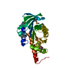
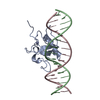
| Entry | Database: PDB / ID: 5ef6 | ||||||
|---|---|---|---|---|---|---|---|
| Title | Structure of HOXB13 complex with methylated DNA | ||||||
 Components Components |
| ||||||
 Keywords Keywords | TRANSCRIPTION / transcription factor / methylated DNA / complex | ||||||
| Function / homology |  Function and homology information Function and homology informationepithelial cell maturation involved in prostate gland development / methyl-CpG binding / regulation of growth / response to testosterone / prostate epithelial cord arborization involved in prostate glandular acinus morphogenesis / epidermis development / response to wounding / DNA-binding transcription repressor activity, RNA polymerase II-specific / sequence-specific double-stranded DNA binding / angiogenesis ...epithelial cell maturation involved in prostate gland development / methyl-CpG binding / regulation of growth / response to testosterone / prostate epithelial cord arborization involved in prostate glandular acinus morphogenesis / epidermis development / response to wounding / DNA-binding transcription repressor activity, RNA polymerase II-specific / sequence-specific double-stranded DNA binding / angiogenesis / transcription regulator complex / sequence-specific DNA binding / DNA-binding transcription factor activity, RNA polymerase II-specific / RNA polymerase II cis-regulatory region sequence-specific DNA binding / regulation of transcription by RNA polymerase II / chromatin / negative regulation of transcription by RNA polymerase II / nucleoplasm Similarity search - Function | ||||||
| Biological species |  Homo sapiens (human) Homo sapiens (human)synthetic construct (others) | ||||||
| Method |  X-RAY DIFFRACTION / SYNCHROTRON / MOLECULAR REPLACEMENT / Resolution: 3 Å X-RAY DIFFRACTION / SYNCHROTRON / MOLECULAR REPLACEMENT / Resolution: 3 Å | ||||||
 Authors Authors | Morgunova, E. / Yin, Y. / Jolma, A. / Popov, A. / Taipale, J. | ||||||
 Citation Citation | Journal: Science / Year: 2017 Title: Impact of cytosine methylation on DNA binding specificities of human transcription factors. Authors: Yin, Y. / Morgunova, E. / Jolma, A. / Kaasinen, E. / Sahu, B. / Khund-Sayeed, S. / Das, P.K. / Kivioja, T. / Dave, K. / Zhong, F. / Nitta, K.R. / Taipale, M. / Popov, A. / Ginno, P.A. / ...Authors: Yin, Y. / Morgunova, E. / Jolma, A. / Kaasinen, E. / Sahu, B. / Khund-Sayeed, S. / Das, P.K. / Kivioja, T. / Dave, K. / Zhong, F. / Nitta, K.R. / Taipale, M. / Popov, A. / Ginno, P.A. / Domcke, S. / Yan, J. / Schubeler, D. / Vinson, C. / Taipale, J. | ||||||
| History |
|
- Structure visualization
| Structure viewer | Molecule: MolmilJmol/JSmol |
|---|
- Downloads & links
-Download
| PDBx/mmCIF format | 5ef6.cif.gz | 143.5 KB | Display | PDBx/mmCIF format |
|---|---|---|---|---|
| PDB format | pdb5ef6.ent.gz | 109.3 KB | Display | PDB format |
| PDBx/mmJSON format | 5ef6.json.gz | Tree view | PDBx/mmJSON format | |
| Others |  Other downloads Other downloads |
-Validation report
| Arichive directory | https://data.pdbj.org/pub/pdb/validation_reports/ef/5ef6ftp://data.pdbj.org/pub/pdb/validation_reports/ef/5ef6 | HTTPS FTP |
|---|
-Related structure data
| Related structure data |  5egoC 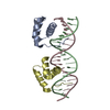 5hodC 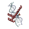 5ltyC  5luxC  5ednS S: Starting model for refinement C: citing same article ( |
|---|---|
| Similar structure data |


-Links
 PDBj
PDBj


- Assembly
| Deposited unit | 
| ||||||||
|---|---|---|---|---|---|---|---|---|---|
| 1 | 
| ||||||||
| 2 | 
| ||||||||
| 3 | 
| ||||||||
| 4 | 
| ||||||||
| Unit cell |
|
-Components
| #1: Protein | Mass: 7510.880 Da / Num. of mol.: 4 / Fragment: UNP residues 217-278 Source method: isolated from a genetically manipulated source Source: (gene. exp.) Homo sapiens (human) / Gene: HOXB13 / Plasmid: pETG20A / Production host:  #2: DNA chain | Mass: 5542.600 Da / Num. of mol.: 4 / Source method: obtained synthetically / Source: (synth.) synthetic construct (others) #3: DNA chain | Mass: 5516.636 Da / Num. of mol.: 4 / Source method: obtained synthetically / Source: (synth.) synthetic construct (others) #4: Water | ChemComp-HOH / |  Mass: 18.015 Da / Num. of mol.: 101 / Source method: isolated from a natural source / Formula: H2O Mass: 18.015 Da / Num. of mol.: 101 / Source method: isolated from a natural source / Formula: H2O |
|---|
-Experimental details
-Experiment
| Experiment | Method: X-RAY DIFFRACTION / Number of used crystals: 1 |
|---|
- Sample preparation
| Crystal | Density Matthews: 2.91 Å3/Da / Density % sol: 57.69 % |
|---|---|
| Crystal grow | Temperature: 293 K / Method: vapor diffusion, sitting drop / pH: 8 Details: PEG 3350, potassium chloride, magnesium chloride, 2-methyl-1-propanol, Tris PH range: 8 |
-Data collection
| Diffraction | Mean temperature: 100 K | ||||||||||||||||||||||||||||||
|---|---|---|---|---|---|---|---|---|---|---|---|---|---|---|---|---|---|---|---|---|---|---|---|---|---|---|---|---|---|---|---|
| Diffraction source | Source: SYNCHROTRON / Site: ESRF  / Beamline: ID23-1 / Wavelength: 0.97242 Å / Beamline: ID23-1 / Wavelength: 0.97242 Å | ||||||||||||||||||||||||||||||
| Detector | Type: PSI PILATUS 6M / Detector: PIXEL / Date: May 26, 2015 | ||||||||||||||||||||||||||||||
| Radiation | Monochromator: Si(111) crystal / Protocol: SINGLE WAVELENGTH / Monochromatic (M) / Laue (L): M / Scattering type: x-ray | ||||||||||||||||||||||||||||||
| Radiation wavelength | Wavelength: 0.97242 Å / Relative weight: 1 | ||||||||||||||||||||||||||||||
| Reflection | Resolution: 2.98→55.16 Å / Num. all: 17811 / Num. obs: 17019 / % possible obs: 97.9 % / Redundancy: 3.3 % / Biso Wilson estimate: 36.6 Å2 / CC1/2: 0.988 / Rmerge(I) obs: 0.202 / Rpim(I) all: 0.13 / Rrim(I) all: 0.241 / Net I/σ(I): 5 / Num. measured all: 57890 | ||||||||||||||||||||||||||||||
| Reflection shell | Diffraction-ID: 1 / Rejects: _
|
- Processing
| Software |
| |||||||||||||||||||||||||||||||||||||||||||||||||||||||||||||||||||||||||||||||||||||||||||
|---|---|---|---|---|---|---|---|---|---|---|---|---|---|---|---|---|---|---|---|---|---|---|---|---|---|---|---|---|---|---|---|---|---|---|---|---|---|---|---|---|---|---|---|---|---|---|---|---|---|---|---|---|---|---|---|---|---|---|---|---|---|---|---|---|---|---|---|---|---|---|---|---|---|---|---|---|---|---|---|---|---|---|---|---|---|---|---|---|---|---|---|---|
| Refinement | Method to determine structure: MOLECULAR REPLACEMENT Starting model: 5edn Resolution: 3→38.169 Å / SU ML: 0.43 / Cross valid method: FREE R-VALUE / σ(F): 1.05 / Phase error: 40.45 / Stereochemistry target values: ML
| |||||||||||||||||||||||||||||||||||||||||||||||||||||||||||||||||||||||||||||||||||||||||||
| Solvent computation | Shrinkage radii: 0.9 Å / VDW probe radii: 1.11 Å / Solvent model: FLAT BULK SOLVENT MODEL | |||||||||||||||||||||||||||||||||||||||||||||||||||||||||||||||||||||||||||||||||||||||||||
| Displacement parameters | Biso max: 145.57 Å2 / Biso mean: 41.4815 Å2 / Biso min: 4.41 Å2 | |||||||||||||||||||||||||||||||||||||||||||||||||||||||||||||||||||||||||||||||||||||||||||
| Refinement step | Cycle: final / Resolution: 3→38.169 Å
| |||||||||||||||||||||||||||||||||||||||||||||||||||||||||||||||||||||||||||||||||||||||||||
| Refine LS restraints |
| |||||||||||||||||||||||||||||||||||||||||||||||||||||||||||||||||||||||||||||||||||||||||||
| LS refinement shell | Refine-ID: X-RAY DIFFRACTION / Total num. of bins used: 12
|
