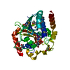
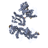
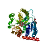
Journal: Sci Rep / Year: 2017 Title: Visualisation of a flexible modular structure of the ER folding-sensor enzyme UGGT. Authors: Tadashi Satoh / Chihong Song / Tong Zhu / Takayasu Toshimori / Kazuyoshi Murata / Yugo Hayashi / Hironari Kamikubo / Takayuki Uchihashi / Koichi Kato / Abstract: In the endoplasmic reticulum (ER), a protein quality control system facilitates the efficient folding of newly synthesised proteins. In this system, a series of N-linked glycan intermediates ...In the endoplasmic reticulum (ER), a protein quality control system facilitates the efficient folding of newly synthesised proteins. In this system, a series of N-linked glycan intermediates displayed on the protein surface serve as quality tags. The ER folding-sensor enzyme UDP-glucose:glycoprotein glucosyltransferase (UGGT) acts as a gatekeeper in the ER quality control system by specifically catalysing monoglucosylation onto incompletely folded glycoproteins, thereby enabling them to interact with lectin-chaperone complexes. Here we characterise the dynamic structure of this enzyme. Our crystallographic data demonstrate that the sensor region is composed of four thioredoxin-like domains followed by a β-rich domain, which are arranged into a C-shaped structure with a large central cavity, while the C-terminal catalytic domain undergoes a ligand-dependent conformational alteration. Furthermore, small-angle X-ray scattering, cryo-electron microscopy and high-speed atomic force microscopy have demonstrated that UGGT has a flexible modular structure in which the smaller catalytic domain is tethered to the larger folding-sensor region with variable spatial arrangements. These findings provide structural insights into the working mechanism whereby UGGT operates as a folding-sensor against a variety of glycoprotein substrates through its flexible modular structure possessing extended hydrophobic surfaces for the recognition of unfolded substrates.
Resolution: 1.4→1.48 Å / Redundancy: 7 % / Rmerge(I) obs: 0.6 / Mean I/σ(I) obs: 2.6 / CC1/2: 0.834 / % possible all: 100
-
Processing
Software
Name
Version
Classification
REFMAC
5.8.0155
refinement
iMOSFLM
datareduction
SCALA
datascaling
SHELXCD
phasing
Refinement
Method to determine structure: SAD / Resolution: 1.4→20 Å / Cor.coef. Fo:Fc: 0.973 / Cor.coef. Fo:Fc free: 0.964 / SU B: 2.074 / SU ML: 0.037 / Cross valid method: THROUGHOUT / ESU R: 0.06 / ESU R Free: 0.055 / Details: HYDROGENS HAVE BEEN ADDED IN THE RIDING POSITIONS
Rfactor
Num. reflection
% reflection
Selection details
Rfree
0.174
2756
4.9 %
RANDOM
Rwork
0.142
-
-
-
obs
0.144
53176
99.9 %
-
Solvent computation
Ion probe radii: 0.8 Å / Shrinkage radii: 0.8 Å / VDW probe radii: 1.2 Å
 Movie
Movie Controller

 Yorodumi
 Open data
 Basic information
 Components
 Keywords
 Function and homology information
 Thermomyces dupontii (fungus)
 X-RAY DIFFRACTION /
  Authors
 Japan, 3items
  Citation
 Structure visualization
 Downloads & links
 Other downloads
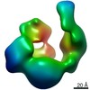
 PDBj
PDBj

 Assembly


 Mass: 566.302 Da / Num. of mol.: 1 / Source method: obtained synthetically / Formula: C15H24N2O17P2
 Mass: 40.078 Da / Num. of mol.: 1 / Source method: obtained synthetically / Formula: Ca
 Mass: 92.094 Da / Num. of mol.: 1 / Source method: obtained synthetically / Formula: C3H8O3
 Mass: 18.015 Da / Num. of mol.: 263 / Source method: isolated from a natural source / Formula: H2O
 Sample preparation
 Processing