 Movie
Movie Controller

[English] 日本語
 Yorodumi
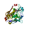
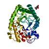
- PDB-5e9w: Crystal structure of mRNA cap guanine-N7 methyltransferase obtain... -
+ Open data

- Basic information
| Entry | Database: PDB / ID: 5e9w | ||||||
|---|---|---|---|---|---|---|---|
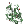
| Title | Crystal structure of mRNA cap guanine-N7 methyltransferase obtained by limited proteolysis | ||||||
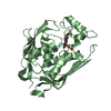
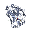
 Components Components | mRNA cap guanine-N7 methyltransferase | ||||||
 Keywords Keywords | TRANSFERASE / mRNA capping | ||||||
| Function / homology |  Function and homology information Function and homology informationmRNA cap methyltransferase RNMT:RAMAC complex / mRNA capping enzyme complex / RNA Pol II CTD phosphorylation and interaction with CE during HIV infection / RNA Pol II CTD phosphorylation and interaction with CE / mRNA Capping / 7-methylguanosine mRNA capping / cellular response to leukemia inhibitory factor / fibrillar center / mRNA (guanine-N7)-methyltransferase / mRNA 5'-cap (guanine-N7-)-methyltransferase activity ...mRNA cap methyltransferase RNMT:RAMAC complex / mRNA capping enzyme complex / RNA Pol II CTD phosphorylation and interaction with CE during HIV infection / RNA Pol II CTD phosphorylation and interaction with CE / mRNA Capping / 7-methylguanosine mRNA capping / cellular response to leukemia inhibitory factor / fibrillar center / mRNA (guanine-N7)-methyltransferase / mRNA 5'-cap (guanine-N7-)-methyltransferase activity / signaling receptor complex / RNA binding / nucleoplasm / nucleus Similarity search - Function | ||||||
| Biological species |  Homo sapiens (human) Homo sapiens (human) | ||||||
| Method |  X-RAY DIFFRACTION / SYNCHROTRON / MOLECULAR REPLACEMENT / Resolution: 2.283 Å X-RAY DIFFRACTION / SYNCHROTRON / MOLECULAR REPLACEMENT / Resolution: 2.283 Å | ||||||
 Authors Authors | Petit, P. / Cowling, V.H. | ||||||
 Citation Citation | Journal: Nucleic Acids Res. / Year: 2016 Title: Molecular basis of RNA guanine-7 methyltransferase (RNMT) activation by RAM. Authors: Varshney, D. / Petit, A.P. / Bueren-Calabuig, J.A. / Jansen, C. / Fletcher, D.A. / Peggie, M. / Weidlich, S. / Scullion, P. / Pisliakov, A.V. / Cowling, V.H. | ||||||
| History |
|
- Structure visualization
| Structure viewer | Molecule: MolmilJmol/JSmol |
|---|
- Downloads & links
-Download
| PDBx/mmCIF format | 5e9w.cif.gz | 463.4 KB | Display | PDBx/mmCIF format |
|---|---|---|---|---|
| PDB format | pdb5e9w.ent.gz | 380.8 KB | Display | PDB format |
| PDBx/mmJSON format | 5e9w.json.gz | Tree view | PDBx/mmJSON format | |
| Others |  Other downloads Other downloads |
-Validation report
| Arichive directory | https://data.pdbj.org/pub/pdb/validation_reports/e9/5e9wftp://data.pdbj.org/pub/pdb/validation_reports/e9/5e9w | HTTPS FTP |
|---|
-Related structure data
| Related structure data |  5e8jC  5e9jC  3bgvS C: citing same article ( S: Starting model for refinement |
|---|---|
| Similar structure data |


-Links
 PDBj
PDBj


- Assembly
| Deposited unit | 
| ||||||||
|---|---|---|---|---|---|---|---|---|---|
| 1 | 
| ||||||||
| 2 | 
| ||||||||
| 3 | 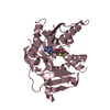
| ||||||||
| 4 | 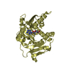
| ||||||||
| Unit cell |
|
-Components
| #1: Protein | Mass: 36716.160 Da / Num. of mol.: 4 / Fragment: UNP residues 165-476 Source method: isolated from a genetically manipulated source Source: (gene. exp.) Homo sapiens (human) / Gene: RNMT, KIAA0398 / Plasmid: PET15 / Production host:  References: UniProt: O43148, mRNA (guanine-N7)-methyltransferase #2: Chemical | ChemComp-SAH /   Mass: 384.411 Da / Num. of mol.: 4 / Source method: obtained synthetically / Formula: C14H20N6O5S Mass: 384.411 Da / Num. of mol.: 4 / Source method: obtained synthetically / Formula: C14H20N6O5S#3: Water | ChemComp-HOH / |  Mass: 18.015 Da / Num. of mol.: 307 / Source method: isolated from a natural source / Formula: H2O Mass: 18.015 Da / Num. of mol.: 307 / Source method: isolated from a natural source / Formula: H2OHas protein modification | N | |
|---|
-Experimental details
-Experiment
| Experiment | Method: X-RAY DIFFRACTION |
|---|
- Sample preparation
| Crystal | Density Matthews: 2.19 Å3/Da / Density % sol: 43.87 % |
|---|---|
| Crystal grow | Temperature: 291 K / Method: vapor diffusion, sitting drop / Details: MES pH 6.5 and 25% PEG 4000, 1/5000 thermolysin |
-Data collection
| Diffraction | Mean temperature: 100 K |
|---|---|
| Diffraction source | Source: SYNCHROTRON / Site: Diamond  / Beamline: I04 / Wavelength: 0.979 Å / Beamline: I04 / Wavelength: 0.979 Å |
| Detector | Type: DECTRIS PILATUS 6M-F / Detector: PIXEL / Date: May 14, 2014 |
| Radiation | Monochromator: Si(111) / Protocol: SINGLE WAVELENGTH / Monochromatic (M) / Laue (L): M / Scattering type: x-ray |
| Radiation wavelength | Wavelength: 0.979 Å / Relative weight: 1 |
| Reflection | Resolution: 2.28→29.58 Å / Num. obs: 57298 / % possible obs: 97 % / Redundancy: 2.8 % / Rmerge(I) obs: 0.08 / Rsym value: 0.07 / Net I/σ(I): 10.3 |
| Reflection shell | Resolution: 2.28→2.41 Å / Redundancy: 2.8 % / Rmerge(I) obs: 0.27 / Mean I/σ(I) obs: 3.9 / % possible all: 97.6 |
- Processing
| Software |
| ||||||||||||||||||||||||||||||||||||||||||||||||||||||||||||||||||||||||||||||||||||||||||||||||||||||||||||||||||||||||||||||||||||||||||||||||||||||||||
|---|---|---|---|---|---|---|---|---|---|---|---|---|---|---|---|---|---|---|---|---|---|---|---|---|---|---|---|---|---|---|---|---|---|---|---|---|---|---|---|---|---|---|---|---|---|---|---|---|---|---|---|---|---|---|---|---|---|---|---|---|---|---|---|---|---|---|---|---|---|---|---|---|---|---|---|---|---|---|---|---|---|---|---|---|---|---|---|---|---|---|---|---|---|---|---|---|---|---|---|---|---|---|---|---|---|---|---|---|---|---|---|---|---|---|---|---|---|---|---|---|---|---|---|---|---|---|---|---|---|---|---|---|---|---|---|---|---|---|---|---|---|---|---|---|---|---|---|---|---|---|---|---|---|---|---|
| Refinement | Method to determine structure: MOLECULAR REPLACEMENT Starting model: 3BGV Resolution: 2.283→29.578 Å / SU ML: 0.25 / Cross valid method: FREE R-VALUE / σ(F): 1.35 / Phase error: 29.64 / Stereochemistry target values: ML
| ||||||||||||||||||||||||||||||||||||||||||||||||||||||||||||||||||||||||||||||||||||||||||||||||||||||||||||||||||||||||||||||||||||||||||||||||||||||||||
| Solvent computation | Shrinkage radii: 0.9 Å / VDW probe radii: 1.11 Å / Solvent model: FLAT BULK SOLVENT MODEL | ||||||||||||||||||||||||||||||||||||||||||||||||||||||||||||||||||||||||||||||||||||||||||||||||||||||||||||||||||||||||||||||||||||||||||||||||||||||||||
| Refinement step | Cycle: LAST / Resolution: 2.283→29.578 Å
| ||||||||||||||||||||||||||||||||||||||||||||||||||||||||||||||||||||||||||||||||||||||||||||||||||||||||||||||||||||||||||||||||||||||||||||||||||||||||||
| Refine LS restraints |
| ||||||||||||||||||||||||||||||||||||||||||||||||||||||||||||||||||||||||||||||||||||||||||||||||||||||||||||||||||||||||||||||||||||||||||||||||||||||||||
| LS refinement shell |
| ||||||||||||||||||||||||||||||||||||||||||||||||||||||||||||||||||||||||||||||||||||||||||||||||||||||||||||||||||||||||||||||||||||||||||||||||||||||||||
| Refinement TLS params. | Method: refined / Origin x: 58.1387 Å / Origin y: 64.3557 Å / Origin z: 62.7484 Å
| ||||||||||||||||||||||||||||||||||||||||||||||||||||||||||||||||||||||||||||||||||||||||||||||||||||||||||||||||||||||||||||||||||||||||||||||||||||||||||
| Refinement TLS group | Selection details: all |
