 Movie
Movie Controller
 Structure viewers
 About Yorodumi Papers


+Search query
-Structure paper
| Title | Visualisation of a flexible modular structure of the ER folding-sensor enzyme UGGT. |
|---|---|
| Journal, issue, pages | Sci Rep, Vol. 7, Issue 1, Page 12142, Year 2017 |
| Publish date | Sep 22, 2017 |
 Authors Authors | Tadashi Satoh / Chihong Song / Tong Zhu / Takayasu Toshimori / Kazuyoshi Murata / Yugo Hayashi / Hironari Kamikubo / Takayuki Uchihashi / Koichi Kato /  |
| PubMed Abstract | In the endoplasmic reticulum (ER), a protein quality control system facilitates the efficient folding of newly synthesised proteins. In this system, a series of N-linked glycan intermediates ...In the endoplasmic reticulum (ER), a protein quality control system facilitates the efficient folding of newly synthesised proteins. In this system, a series of N-linked glycan intermediates displayed on the protein surface serve as quality tags. The ER folding-sensor enzyme UDP-glucose:glycoprotein glucosyltransferase (UGGT) acts as a gatekeeper in the ER quality control system by specifically catalysing monoglucosylation onto incompletely folded glycoproteins, thereby enabling them to interact with lectin-chaperone complexes. Here we characterise the dynamic structure of this enzyme. Our crystallographic data demonstrate that the sensor region is composed of four thioredoxin-like domains followed by a β-rich domain, which are arranged into a C-shaped structure with a large central cavity, while the C-terminal catalytic domain undergoes a ligand-dependent conformational alteration. Furthermore, small-angle X-ray scattering, cryo-electron microscopy and high-speed atomic force microscopy have demonstrated that UGGT has a flexible modular structure in which the smaller catalytic domain is tethered to the larger folding-sensor region with variable spatial arrangements. These findings provide structural insights into the working mechanism whereby UGGT operates as a folding-sensor against a variety of glycoprotein substrates through its flexible modular structure possessing extended hydrophobic surfaces for the recognition of unfolded substrates. |
 External links External links | Sci Rep / PubMed:28939828 / PubMed Central |
| Methods | EM (single particle) / X-ray diffraction |
| Resolution | 1.35 - 22.9 Å |
| Structure data | 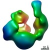 EMDB-30386: 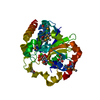 PDB-5h18: 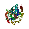 PDB-5y7f: 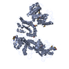 PDB-5y7o: |
| Chemicals |  ChemComp-UPG:  ChemComp-CA:  ChemComp-GOL:  ChemComp-HOH:  ChemComp-UDP:  ChemComp-TRS: |
| Source |
|
 Keywords Keywords | TRANSFERASE / ENDOPLASMIC RETICULUM / QUALITY CONTROL / GLUCOSYLTRANSFERASE / FOLDING SENSOR |

 thermomyces dupontii (fungus)