 Movie
Movie Controller


+ Open data

- Basic information
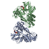
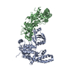
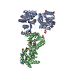
| Entry | Database: PDB / ID: 5gru | |||||||||
|---|---|---|---|---|---|---|---|---|---|---|
| Title | Structure of mono-specific diabody | |||||||||
 Components Components |
| |||||||||
 Keywords Keywords | IMMUNE SYSTEM / diabody / antibody fragment | |||||||||
| Function / homology |  Function and homology information Function and homology informationdetection of maltose stimulus / maltose transport complex / carbohydrate transport / carbohydrate transmembrane transporter activity / maltose binding / maltose transport / maltodextrin transmembrane transport / ATP-binding cassette (ABC) transporter complex, substrate-binding subunit-containing / ATP-binding cassette (ABC) transporter complex / cell chemotaxis ...detection of maltose stimulus / maltose transport complex / carbohydrate transport / carbohydrate transmembrane transporter activity / maltose binding / maltose transport / maltodextrin transmembrane transport / ATP-binding cassette (ABC) transporter complex, substrate-binding subunit-containing / ATP-binding cassette (ABC) transporter complex / cell chemotaxis / outer membrane-bounded periplasmic space / periplasmic space / DNA damage response / membrane Similarity search - Function | |||||||||
| Biological species |   Homo sapiens (human) Homo sapiens (human) | |||||||||
| Method |  X-RAY DIFFRACTION / SYNCHROTRON / MOLECULAR REPLACEMENT / Resolution: 1.955 Å X-RAY DIFFRACTION / SYNCHROTRON / MOLECULAR REPLACEMENT / Resolution: 1.955 Å | |||||||||
 Authors Authors | Kim, J.H. / Song, D.H. / Youn, S.J. / Cho, G. / Lee, H. / Lee, J.O. | |||||||||
| Funding support |  Korea, Republic Of, 2items Korea, Republic Of, 2items
| |||||||||
 Citation Citation | Journal: Sci Rep / Year: 2016 Title: Crystal structure of mono- and bi-specific diabodies and reduction of their structural flexibility by introduction of disulfide bridges at the Fv interface. Authors: Kim, J.H. / Song, D.H. / Youn, S.J. / Kim, J.W. / Cho, G. / Kim, S.C. / Lee, H. / Jin, M.S. / Lee, J.O. | |||||||||
| History |
|
- Structure visualization
| Structure viewer | Molecule: MolmilJmol/JSmol |
|---|
- Downloads & links
-Download
| PDBx/mmCIF format | 5gru.cif.gz | 191 KB | Display | PDBx/mmCIF format |
|---|---|---|---|---|
| PDB format | pdb5gru.ent.gz | 147.6 KB | Display | PDB format |
| PDBx/mmJSON format | 5gru.json.gz | Tree view | PDBx/mmJSON format | |
| Others |  Other downloads Other downloads |
-Validation report
| Arichive directory | https://data.pdbj.org/pub/pdb/validation_reports/gr/5gruftp://data.pdbj.org/pub/pdb/validation_reports/gr/5gru | HTTPS FTP |
|---|
-Related structure data
| Related structure data | 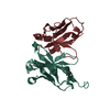 5grvC 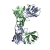 5grwC 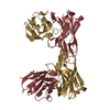 5grxC 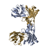 5gryC 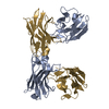 5grzC 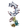 5gs0C  5gs1C 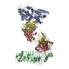 5gs2C 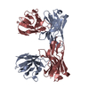 5gs3C  1ezvS 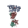 3pgfS C: citing same article ( S: Starting model for refinement |
|---|---|
| Similar structure data |


-Links
 PDBj
PDBj


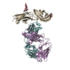
- Assembly
| Deposited unit | 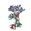
| ||||||||
|---|---|---|---|---|---|---|---|---|---|
| 1 |
| ||||||||
| Unit cell |
|
-Components
| #1: Protein | Mass: 42147.621 Da / Num. of mol.: 1 / Fragment: UNP RESIDUES 27-392 Source method: isolated from a genetically manipulated source Source: (gene. exp.) |
|---|---|
| #2: Antibody | Mass: 26993.654 Da / Num. of mol.: 1 Source method: isolated from a genetically manipulated source Source: (gene. exp.) Homo sapiens (human) / Production host:  Trichoplusia ni (cabbage looper) Trichoplusia ni (cabbage looper) |
| #3: Antibody | Mass: 24800.551 Da / Num. of mol.: 1 Source method: isolated from a genetically manipulated source Source: (gene. exp.) Trichoplusia ni (cabbage looper) |
| #4: Water | ChemComp-HOH /  Mass: 18.015 Da / Num. of mol.: 809 / Source method: isolated from a natural source / Formula: H2O Mass: 18.015 Da / Num. of mol.: 809 / Source method: isolated from a natural source / Formula: H2O |
| Has protein modification | Y |
-Experimental details
-Experiment
| Experiment | Method: X-RAY DIFFRACTION / Number of used crystals: 1 |
|---|
- Sample preparation
| Crystal | Density Matthews: 2.72 Å3/Da / Density % sol: 54.75 % |
|---|---|
| Crystal grow | Temperature: 296 K / Method: vapor diffusion, sitting drop / pH: 5.6 Details: 0.1 M Sodium citrate tribasic pH 5.6, 27.5% PEG 12000, 0.1 M Sodium iodide |
-Data collection
| Diffraction | Mean temperature: 80 K |
|---|---|
| Diffraction source | Source: SYNCHROTRON / Site: PAL/PLS / Beamline: 7A (6B, 6C1) / Wavelength: 0.97933 Å |
| Detector | Type: ADSC QUANTUM 270 / Detector: CCD / Date: Sep 14, 2013 |
| Radiation | Protocol: SINGLE WAVELENGTH / Monochromatic (M) / Laue (L): M / Scattering type: x-ray |
| Radiation wavelength | Wavelength: 0.97933 Å / Relative weight: 1 |
| Reflection | Resolution: 1.95→50 Å / Num. obs: 70116 / % possible obs: 98.9 % / Redundancy: 3.2 % / Net I/σ(I): 14.2 |
| Reflection shell | Resolution: 1.95→2.02 Å |
- Processing
| Software |
| |||||||||||||||||||||||||||||||||||||||||||||||||||||||||||||||||||||||||||||||||||||||||||||||||||||||||
|---|---|---|---|---|---|---|---|---|---|---|---|---|---|---|---|---|---|---|---|---|---|---|---|---|---|---|---|---|---|---|---|---|---|---|---|---|---|---|---|---|---|---|---|---|---|---|---|---|---|---|---|---|---|---|---|---|---|---|---|---|---|---|---|---|---|---|---|---|---|---|---|---|---|---|---|---|---|---|---|---|---|---|---|---|---|---|---|---|---|---|---|---|---|---|---|---|---|---|---|---|---|---|---|---|---|---|
| Refinement | Method to determine structure: MOLECULAR REPLACEMENT Starting model: 1EZV, 3PGF Resolution: 1.955→33.779 Å / SU ML: 0.22 / Cross valid method: FREE R-VALUE / σ(F): 1.39 / Phase error: 23.34
| |||||||||||||||||||||||||||||||||||||||||||||||||||||||||||||||||||||||||||||||||||||||||||||||||||||||||
| Solvent computation | Shrinkage radii: 0.9 Å / VDW probe radii: 1.11 Å | |||||||||||||||||||||||||||||||||||||||||||||||||||||||||||||||||||||||||||||||||||||||||||||||||||||||||
| Refinement step | Cycle: LAST / Resolution: 1.955→33.779 Å
| |||||||||||||||||||||||||||||||||||||||||||||||||||||||||||||||||||||||||||||||||||||||||||||||||||||||||
| Refine LS restraints |
| |||||||||||||||||||||||||||||||||||||||||||||||||||||||||||||||||||||||||||||||||||||||||||||||||||||||||
| LS refinement shell |
|
