 Movie
Movie Controller

[English] 日本語
 Yorodumi
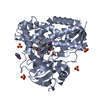
- PDB-5g6t: Crystal structure of Zn-containing NagZ H174A mutant from Pseudom... -
+ Open data

- Basic information
| Entry | Database: PDB / ID: 5g6t | ||||||
|---|---|---|---|---|---|---|---|
| Title | Crystal structure of Zn-containing NagZ H174A mutant from Pseudomonas aeruginosa | ||||||
 Components Components | BETA-HEXOSAMINIDASE | ||||||
 Keywords Keywords | HYDROLASE / CELL-WALL RECYCLING / ANTIBIOTIC RESISTANCE / GLYCOSIDE HYDROLASE / N-ACETYLGLUCOSAMINIDASE / BETA-HEXOSAMINIDASE / PEPTIDOGLYCAN | ||||||
| Function / homology |  Function and homology information Function and homology informationbeta-N-acetylhexosaminidase / peptidoglycan turnover / peptidoglycan biosynthetic process / beta-N-acetylglucosaminidase activity / cell wall organization / regulation of cell shape / carbohydrate metabolic process / cell division / response to antibiotic / cytosol Similarity search - Function | ||||||
| Biological species |   PSEUDOMONAS AERUGINOSA (bacteria) PSEUDOMONAS AERUGINOSA (bacteria) | ||||||
| Method |  X-RAY DIFFRACTION / SYNCHROTRON / MOLECULAR REPLACEMENT / Resolution: 2.15 Å X-RAY DIFFRACTION / SYNCHROTRON / MOLECULAR REPLACEMENT / Resolution: 2.15 Å | ||||||
 Authors Authors | Acebron, I. / Artola-Recolons, C. / Mahasenan, K. / Mobashery, S. / Hermoso, J.A. | ||||||
 Citation Citation | Journal: J. Am. Chem. Soc. / Year: 2017 Title: Catalytic Cycle of the N-Acetylglucosaminidase NagZ from Pseudomonas aeruginosa. Authors: Acebron, I. / Mahasenan, K.V. / De Benedetti, S. / Lee, M. / Artola-Recolons, C. / Hesek, D. / Wang, H. / Hermoso, J.A. / Mobashery, S. | ||||||
| History |
|
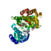
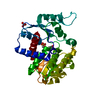
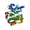
- Structure visualization
| Structure viewer | Molecule: MolmilJmol/JSmol |
|---|
- Downloads & links
-Download
| PDBx/mmCIF format | 5g6t.cif.gz | 264 KB | Display | PDBx/mmCIF format |
|---|---|---|---|---|
| PDB format | pdb5g6t.ent.gz | 214.5 KB | Display | PDB format |
| PDBx/mmJSON format | 5g6t.json.gz | Tree view | PDBx/mmJSON format | |
| Others |  Other downloads Other downloads |
-Validation report
| Arichive directory | https://data.pdbj.org/pub/pdb/validation_reports/g6/5g6tftp://data.pdbj.org/pub/pdb/validation_reports/g6/5g6t | HTTPS FTP |
|---|
-Related structure data
| Related structure data |  5g1mC  5g2mC  5g3rC  5g5kC  5g5uC  5ly7C C: citing same article ( |
|---|---|
| Similar structure data |


-Links
 PDBj
PDBj- Assembly
| Deposited unit | 
| ||||||||
|---|---|---|---|---|---|---|---|---|---|
| 1 | 
| ||||||||
| 2 | 
| ||||||||
| Unit cell |
|
-Components
| #1: Protein | Mass: 38250.605 Da / Num. of mol.: 2 / Mutation: YES Source method: isolated from a genetically manipulated source Details: BOTH PROTEIN CHAINS CONTAIN A HIS RESIDUE AT POSITION -1 FROM THE FUSION TAG USED FOR PURIFICATION. THE SEQUENCE INCLUDES THE SINGLE-POINT MUTATION H174A THAT MAKES THIS PROTEIN INACTIVE. Source: (gene. exp.) PSEUDOMONAS AERUGINOSA (bacteria) / Strain: PAO1 / Production host: #2: Chemical |   Mass: 65.409 Da / Num. of mol.: 2 / Source method: obtained synthetically / Formula: Zn Mass: 65.409 Da / Num. of mol.: 2 / Source method: obtained synthetically / Formula: Zn#3: Chemical | ChemComp-PEG / |   Mass: 106.120 Da / Num. of mol.: 1 / Source method: obtained synthetically / Formula: C4H10O3 Mass: 106.120 Da / Num. of mol.: 1 / Source method: obtained synthetically / Formula: C4H10O3#4: Water | ChemComp-HOH / |  Mass: 18.015 Da / Num. of mol.: 393 / Source method: isolated from a natural source / Formula: H2O Mass: 18.015 Da / Num. of mol.: 393 / Source method: isolated from a natural source / Formula: H2ONonpolymer details | ZINC ION (ZN): THE SOURCE OF ZN IS UNKNOWN. IT IS LIKELY THAT ZN COMES EITHER THE EXPRESSION HOST ...ZINC ION (ZN): THE SOURCE OF ZN IS UNKNOWN. IT IS LIKELY THAT ZN COMES EITHER THE EXPRESSION | Sequence details | THE EXPERIMENT | |
|---|
-Experimental details
-Experiment
| Experiment | Method: X-RAY DIFFRACTION / Number of used crystals: 1 |
|---|
- Sample preparation
| Crystal | Density Matthews: 2.07 Å3/Da / Density % sol: 40.8 % / Description: NONE |
|---|---|
| Crystal grow | pH: 6 Details: 30% PEG 8000 100 MM SODIUM CACODYLATE PH 6.0 200 MM SODIUM ACETATE PH 6.0 |
-Data collection
| Diffraction | Mean temperature: 100 K |
|---|---|
| Diffraction source | Source: SYNCHROTRON / Site: ALBA  / Beamline: XALOC / Wavelength: 0.97948 / Beamline: XALOC / Wavelength: 0.97948 |
| Detector | Type: DECTRIS PILATUS 6M / Detector: PIXEL / Date: Oct 17, 2015 / Details: KB MIRRORS |
| Radiation | Monochromator: SI(111) CHANNEL-CUT / Protocol: SINGLE WAVELENGTH / Monochromatic (M) / Laue (L): M / Scattering type: x-ray |
| Radiation wavelength | Wavelength: 0.97948 Å / Relative weight: 1 |
| Reflection | Resolution: 2.15→47.68 Å / Num. obs: 36891 / % possible obs: 97.2 % / Observed criterion σ(I): 1.5 / Redundancy: 4.5 % / Biso Wilson estimate: 24.01 Å2 / Rmerge(I) obs: 0.18 / Net I/σ(I): 8.3 |
| Reflection shell | Resolution: 2.15→2.22 Å / Redundancy: 4.4 % / Rmerge(I) obs: 0.95 / Mean I/σ(I) obs: 1.6 / % possible all: 97.3 |
- Processing
| Software |
| |||||||||||||||||||||||||||||||||||||||||||||||||||||||||||||||||||||||||||||||||||||||||||||||||||||||||||||||||||||||||||||||||||||||||||||||||||||||||||||||||||||||||||||||||||||||||||||||||||||||||||||||||||||||||||||||||
|---|---|---|---|---|---|---|---|---|---|---|---|---|---|---|---|---|---|---|---|---|---|---|---|---|---|---|---|---|---|---|---|---|---|---|---|---|---|---|---|---|---|---|---|---|---|---|---|---|---|---|---|---|---|---|---|---|---|---|---|---|---|---|---|---|---|---|---|---|---|---|---|---|---|---|---|---|---|---|---|---|---|---|---|---|---|---|---|---|---|---|---|---|---|---|---|---|---|---|---|---|---|---|---|---|---|---|---|---|---|---|---|---|---|---|---|---|---|---|---|---|---|---|---|---|---|---|---|---|---|---|---|---|---|---|---|---|---|---|---|---|---|---|---|---|---|---|---|---|---|---|---|---|---|---|---|---|---|---|---|---|---|---|---|---|---|---|---|---|---|---|---|---|---|---|---|---|---|---|---|---|---|---|---|---|---|---|---|---|---|---|---|---|---|---|---|---|---|---|---|---|---|---|---|---|---|---|---|---|---|---|---|---|---|---|---|---|---|---|---|---|---|---|---|---|---|---|
| Refinement | Method to determine structure: MOLECULAR REPLACEMENT Starting model: APO STRUCTURE OF NAGZ FROM PSEUDOMONAS AERUGINOSA Resolution: 2.15→44.937 Å / SU ML: 0.26 / σ(F): 1.35 / Phase error: 24.87 / Stereochemistry target values: ML Details: RESIDUES 124-135 AND 171-174 IN CHAIN A, AND REIDUES 124-134, 171-175 AND 249-251 IN CHAIN B WERE NOT MODELED DUE TO POOR ELECTRON DENSITY.
| |||||||||||||||||||||||||||||||||||||||||||||||||||||||||||||||||||||||||||||||||||||||||||||||||||||||||||||||||||||||||||||||||||||||||||||||||||||||||||||||||||||||||||||||||||||||||||||||||||||||||||||||||||||||||||||||||
| Solvent computation | Shrinkage radii: 0.9 Å / VDW probe radii: 1.11 Å / Solvent model: FLAT BULK SOLVENT MODEL | |||||||||||||||||||||||||||||||||||||||||||||||||||||||||||||||||||||||||||||||||||||||||||||||||||||||||||||||||||||||||||||||||||||||||||||||||||||||||||||||||||||||||||||||||||||||||||||||||||||||||||||||||||||||||||||||||
| Displacement parameters | Biso mean: 34.47 Å2 | |||||||||||||||||||||||||||||||||||||||||||||||||||||||||||||||||||||||||||||||||||||||||||||||||||||||||||||||||||||||||||||||||||||||||||||||||||||||||||||||||||||||||||||||||||||||||||||||||||||||||||||||||||||||||||||||||
| Refinement step | Cycle: LAST / Resolution: 2.15→44.937 Å
| |||||||||||||||||||||||||||||||||||||||||||||||||||||||||||||||||||||||||||||||||||||||||||||||||||||||||||||||||||||||||||||||||||||||||||||||||||||||||||||||||||||||||||||||||||||||||||||||||||||||||||||||||||||||||||||||||
| Refine LS restraints |
| |||||||||||||||||||||||||||||||||||||||||||||||||||||||||||||||||||||||||||||||||||||||||||||||||||||||||||||||||||||||||||||||||||||||||||||||||||||||||||||||||||||||||||||||||||||||||||||||||||||||||||||||||||||||||||||||||
| LS refinement shell |
| |||||||||||||||||||||||||||||||||||||||||||||||||||||||||||||||||||||||||||||||||||||||||||||||||||||||||||||||||||||||||||||||||||||||||||||||||||||||||||||||||||||||||||||||||||||||||||||||||||||||||||||||||||||||||||||||||
| Refinement TLS params. | Method: refined / Refine-ID: X-RAY DIFFRACTION
| |||||||||||||||||||||||||||||||||||||||||||||||||||||||||||||||||||||||||||||||||||||||||||||||||||||||||||||||||||||||||||||||||||||||||||||||||||||||||||||||||||||||||||||||||||||||||||||||||||||||||||||||||||||||||||||||||
| Refinement TLS group |
|
