 Movie
Movie Controller

[English] 日本語
 Yorodumi
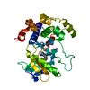
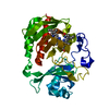
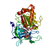
- PDB-2r6h: Crystal structure of the domain comprising the NAD binding and th... -
+ Open data

- Basic information
| Entry | Database: PDB / ID: 2r6h | ||||||
|---|---|---|---|---|---|---|---|
| Title | Crystal structure of the domain comprising the NAD binding and the FAD binding regions of the NADH:ubiquinone oxidoreductase, Na translocating, F subunit from Porphyromonas gingivalis | ||||||
 Components Components | NADH:ubiquinone oxidoreductase, Na translocating, F subunit | ||||||
 Keywords Keywords | OXIDOREDUCTASE / alpha-beta-alpha sandwich / Structural Genomics / PSI-2 / Protein Structure Initiative / Midwest Center for Structural Genomics / MCSG / Iron / Iron-sulfur / Metal-binding / Ubiquinone | ||||||
| Function / homology |  Function and homology information Function and homology informationNADH:ubiquinone reductase (Na+-transporting) / oxidoreductase activity, acting on NAD(P)H, quinone or similar compound as acceptor / sodium ion transport / 2 iron, 2 sulfur cluster binding / electron transfer activity / nucleotide binding / metal ion binding / plasma membrane Similarity search - Function | ||||||
| Biological species |  Porphyromonas gingivalis (bacteria) Porphyromonas gingivalis (bacteria) | ||||||
| Method |  X-RAY DIFFRACTION / SYNCHROTRON / SAD / Resolution: 2.95 Å X-RAY DIFFRACTION / SYNCHROTRON / SAD / Resolution: 2.95 Å | ||||||
 Authors Authors | Kim, Y. / Mulligan, R. / Moy, S. / Joachimiak, A. / Midwest Center for Structural Genomics (MCSG) | ||||||
 Citation Citation | Journal: To be Published Title: Crystal Structure of the Domain Comprising the Regions Binding NAD and FAD from the NADH:Ubiquinone Oxidoreductase, Na Translocating, F Subunit from Porphyromonas gingivalis. Authors: Kim, Y. / Mulligan, R. / Moy, S. / Joachimiak, A. | ||||||
| History |
|
- Structure visualization
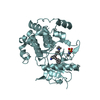
| Structure viewer | Molecule: MolmilJmol/JSmol |
|---|
- Downloads & links
-Download
| PDBx/mmCIF format | 2r6h.cif.gz | 480.2 KB | Display | PDBx/mmCIF format |
|---|---|---|---|---|
| PDB format | pdb2r6h.ent.gz | 402.2 KB | Display | PDB format |
| PDBx/mmJSON format | 2r6h.json.gz | Tree view | PDBx/mmJSON format | |
| Others |  Other downloads Other downloads |
-Validation report
| Arichive directory | https://data.pdbj.org/pub/pdb/validation_reports/r6/2r6hftp://data.pdbj.org/pub/pdb/validation_reports/r6/2r6h | HTTPS FTP |
|---|
-Related structure data
| Similar structure data | |
|---|---|
| Other databases |


-Links
 PDBj
PDBj


- Assembly
| Deposited unit | 
| ||||||||
|---|---|---|---|---|---|---|---|---|---|
| 1 | 
| ||||||||
| 2 | 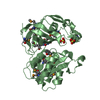
| ||||||||
| 3 | 
| ||||||||
| 4 | 
| ||||||||
| Unit cell |
| ||||||||
| Details | Authors state that the biological unit is unknown. |
-Components
| #1: Protein | Mass: 33819.438 Da / Num. of mol.: 4 / Fragment: The NAD and FAD binding region: Residues 126-412 Source method: isolated from a genetically manipulated source Source: (gene. exp.) Porphyromonas gingivalis (bacteria) / Strain: W83 / Gene: nqrF, PG_2177 / Plasmid: pMCSG7 / Production host: References: UniProt: Q7MT22, Oxidoreductases; Acting on NADH or NADPH; With a quinone or similar compound as acceptor #2: Chemical | ChemComp-SO4 /   Mass: 96.063 Da / Num. of mol.: 11 / Source method: obtained synthetically / Formula: SO4 Mass: 96.063 Da / Num. of mol.: 11 / Source method: obtained synthetically / Formula: SO4#3: Chemical | ChemComp-FAD /   Mass: 785.550 Da / Num. of mol.: 4 / Source method: obtained synthetically / Formula: C27H33N9O15P2 / Comment: FAD*YM Mass: 785.550 Da / Num. of mol.: 4 / Source method: obtained synthetically / Formula: C27H33N9O15P2 / Comment: FAD*YM#4: Water | ChemComp-HOH / |  Mass: 18.015 Da / Num. of mol.: 254 / Source method: isolated from a natural source / Formula: H2O Mass: 18.015 Da / Num. of mol.: 254 / Source method: isolated from a natural source / Formula: H2OHas protein modification | Y | |
|---|
-Experimental details
-Experiment
| Experiment | Method: X-RAY DIFFRACTION / Number of used crystals: 1 |
|---|
- Sample preparation
| Crystal | Density Matthews: 3.48 Å3/Da / Density % sol: 64.69 % |
|---|---|
| Crystal grow | Temperature: 289 K / Method: vapor diffusion, sitting drop / pH: 8.5 Details: 0.1 M Tris-HCl pH 8.5, 2M Ammonium sulfate, VAPOR DIFFUSION, SITTING DROP, temperature 289K |
-Data collection
| Diffraction | Mean temperature: 100 K |
|---|---|
| Diffraction source | Source: SYNCHROTRON / Site: APS  / Beamline: 19-BM / Wavelength: 0.9794 Å / Beamline: 19-BM / Wavelength: 0.9794 Å |
| Detector | Type: SBC-3 / Detector: CCD / Date: Dec 18, 2006 / Details: mirrors |
| Radiation | Monochromator: double crystal / Protocol: SINGLE WAVELENGTH / Monochromatic (M) / Laue (L): M / Scattering type: x-ray |
| Radiation wavelength | Wavelength: 0.9794 Å / Relative weight: 1 |
| Reflection | Resolution: 2.95→47.54 Å / Num. all: 40721 / Num. obs: 40721 / % possible obs: 100 % / Observed criterion σ(F): 0 / Observed criterion σ(I): 0 / Redundancy: 17.5 % / Rsym value: 0.179 / Net I/σ(I): 5.2 |
| Reflection shell | Resolution: 2.95→3.06 Å / Redundancy: 16.8 % / Mean I/σ(I) obs: 3.7 / Num. unique all: 3999 / Rsym value: 0.736 / % possible all: 100 |
- Processing
| Software |
| |||||||||||||||||||||||||
|---|---|---|---|---|---|---|---|---|---|---|---|---|---|---|---|---|---|---|---|---|---|---|---|---|---|---|
| Refinement | Method to determine structure: SAD / Resolution: 2.95→47.54 Å / Cross valid method: THROUGHOUT / σ(F): 0 / σ(I): 0 / Stereochemistry target values: Engh & Huber
| |||||||||||||||||||||||||
| Displacement parameters | Biso mean: 47.411 Å2
| |||||||||||||||||||||||||
| Refinement step | Cycle: LAST / Resolution: 2.95→47.54 Å
| |||||||||||||||||||||||||
| Refine LS restraints |
| |||||||||||||||||||||||||
| LS refinement shell | Resolution: 2.95→3.03 Å
|
