 Movie
Movie Controller


+ Open data

- Basic information
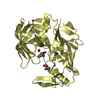
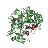
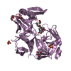
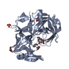
| Entry | Database: PDB / ID: 7agd | ||||||
|---|---|---|---|---|---|---|---|
| Title | Protease Sapp1p from Candida parapsilosis in complex with KB75 | ||||||
 Components Components |
| ||||||
 Keywords Keywords | ANTIBIOTIC / Secreted aspartic protease / virulence factor / candidiasis / peptidomimetic inhibitors | ||||||
| Function / homology |  Function and homology information Function and homology informationcandidapepsin / aspartic-type endopeptidase activity / proteolysis / extracellular region Similarity search - Function | ||||||
| Biological species |  Candida parapsilosis (fungus) Candida parapsilosis (fungus) Streptomyces albus (bacteria) Streptomyces albus (bacteria) | ||||||
| Method |  X-RAY DIFFRACTION / SYNCHROTRON / MOLECULAR REPLACEMENT / Resolution: 1.8 Å X-RAY DIFFRACTION / SYNCHROTRON / MOLECULAR REPLACEMENT / Resolution: 1.8 Å | ||||||
 Authors Authors | Dostal, J. / Heidingsfeld, O. / Brynda, J. | ||||||
| Funding support |  Czech Republic, 1items Czech Republic, 1items
| ||||||
 Citation Citation | Journal: J Enzyme Inhib Med Chem / Year: 2021 Title: Structural determinants for subnanomolar inhibition of the secreted aspartic protease Sapp1p from Candida parapsilosis . Authors: Dostal, J. / Brynda, J. / Vankova, L. / Zia, S.R. / Pichova, I. / Heidingsfeld, O. / Lepsik, M. | ||||||
| History |
|
- Structure visualization
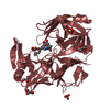
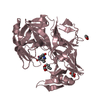
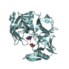
| Structure viewer | Molecule: MolmilJmol/JSmol |
|---|
- Downloads & links
-Download
| PDBx/mmCIF format | 7agd.cif.gz | 286.2 KB | Display | PDBx/mmCIF format |
|---|---|---|---|---|
| PDB format | pdb7agd.ent.gz | 231.4 KB | Display | PDB format |
| PDBx/mmJSON format | 7agd.json.gz | Tree view | PDBx/mmJSON format | |
| Others |  Other downloads Other downloads |
-Validation report
| Arichive directory | https://data.pdbj.org/pub/pdb/validation_reports/ag/7agdftp://data.pdbj.org/pub/pdb/validation_reports/ag/7agd | HTTPS FTP |
|---|
-Related structure data
| Related structure data |  7agbC 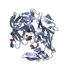 7agcC 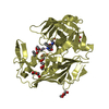 7ageC 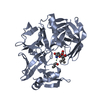 4y9wS S: Starting model for refinement C: citing same article ( |
|---|---|
| Similar structure data |


-Links
 PDBj
PDBj- Assembly
| Deposited unit | 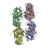
| ||||||||
|---|---|---|---|---|---|---|---|---|---|
| 1 | 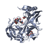
| ||||||||
| 2 | 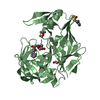
| ||||||||
| 3 | 
| ||||||||
| 4 | 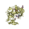
| ||||||||
| Unit cell |
|
-Components
-Protein / Protein/peptide , 2 types, 8 molecules ABCDIJKL
| #1: Protein | Mass: 35865.160 Da / Num. of mol.: 4 Source method: isolated from a genetically manipulated source Source: (gene. exp.) Candida parapsilosis (fungus) / Gene: SAPP1 / Production host: Candida parapsilosis (fungus) / References: UniProt: B8YPM3, candidapepsin#2: Protein/peptide | Mass: 841.002 Da / Num. of mol.: 4 / Source method: obtained synthetically / Source: (synth.) Streptomyces albus (bacteria) |
|---|
-Non-polymers , 7 types, 787 molecules 


| #3: Chemical |  Mass: 238.278 Da / Num. of mol.: 2 / Source method: obtained synthetically / Formula: C10H22O6 / Comment: precipitant*YM Mass: 238.278 Da / Num. of mol.: 2 / Source method: obtained synthetically / Formula: C10H22O6 / Comment: precipitant*YM#4: Chemical | ChemComp-PEG / |  Mass: 106.120 Da / Num. of mol.: 1 / Source method: obtained synthetically / Formula: C4H10O3 Mass: 106.120 Da / Num. of mol.: 1 / Source method: obtained synthetically / Formula: C4H10O3#5: Chemical |  Mass: 150.173 Da / Num. of mol.: 3 / Source method: obtained synthetically / Formula: C6H14O4 Mass: 150.173 Da / Num. of mol.: 3 / Source method: obtained synthetically / Formula: C6H14O4#6: Chemical | ChemComp-MES / |  Mass: 195.237 Da / Num. of mol.: 1 / Source method: obtained synthetically / Formula: C6H13NO4S / Comment: pH buffer*YM Mass: 195.237 Da / Num. of mol.: 1 / Source method: obtained synthetically / Formula: C6H13NO4S / Comment: pH buffer*YM#7: Chemical | ChemComp-NA / |  Mass: 22.990 Da / Num. of mol.: 1 / Source method: obtained synthetically / Formula: Na Mass: 22.990 Da / Num. of mol.: 1 / Source method: obtained synthetically / Formula: Na#8: Chemical | ChemComp-FOR / |  Mass: 30.026 Da / Num. of mol.: 1 / Source method: obtained synthetically / Formula: CH2O Mass: 30.026 Da / Num. of mol.: 1 / Source method: obtained synthetically / Formula: CH2O#9: Water | ChemComp-HOH / | Mass: 18.015 Da / Num. of mol.: 778 / Source method: isolated from a natural source / Formula: H2O |
|---|
-Details
| Has ligand of interest | Y |
|---|
-Experimental details
-Experiment
| Experiment | Method: X-RAY DIFFRACTION / Number of used crystals: 1 |
|---|
- Sample preparation
| Crystal | Density Matthews: 3.68 Å3/Da / Density % sol: 66.6 % |
|---|---|
| Crystal grow | Temperature: 292 K / Method: vapor diffusion, hanging drop Details: 100-fold molar inhibitor excess, Cpr=20mg/ml; drops: 0.002ml protein + 0.001ml reservoir; reservoir: 0.1M MES pH 6.5, 30% v/v PEG 400, VAPOR DIFFUSION, HANGING DROP, temperature 292K |
-Data collection
| Diffraction | Mean temperature: 100 K / Serial crystal experiment: N | |||||||||||||||||||||||||||||||||||||||||||||||||||||||||||||||||||||||||||||||||||||||||||||||||||||||||||||||||||||||||||||||||||||||||||||||||||||||||||||||||||||||||||||||||||||||||||||||||||||||||||||||||||||||||||||||||||||||
|---|---|---|---|---|---|---|---|---|---|---|---|---|---|---|---|---|---|---|---|---|---|---|---|---|---|---|---|---|---|---|---|---|---|---|---|---|---|---|---|---|---|---|---|---|---|---|---|---|---|---|---|---|---|---|---|---|---|---|---|---|---|---|---|---|---|---|---|---|---|---|---|---|---|---|---|---|---|---|---|---|---|---|---|---|---|---|---|---|---|---|---|---|---|---|---|---|---|---|---|---|---|---|---|---|---|---|---|---|---|---|---|---|---|---|---|---|---|---|---|---|---|---|---|---|---|---|---|---|---|---|---|---|---|---|---|---|---|---|---|---|---|---|---|---|---|---|---|---|---|---|---|---|---|---|---|---|---|---|---|---|---|---|---|---|---|---|---|---|---|---|---|---|---|---|---|---|---|---|---|---|---|---|---|---|---|---|---|---|---|---|---|---|---|---|---|---|---|---|---|---|---|---|---|---|---|---|---|---|---|---|---|---|---|---|---|---|---|---|---|---|---|---|---|---|---|---|---|---|---|---|---|---|
| Diffraction source | Source: SYNCHROTRON / Site: BESSY  / Beamline: 14.1 / Wavelength: 0.91841 Å / Beamline: 14.1 / Wavelength: 0.91841 Å | |||||||||||||||||||||||||||||||||||||||||||||||||||||||||||||||||||||||||||||||||||||||||||||||||||||||||||||||||||||||||||||||||||||||||||||||||||||||||||||||||||||||||||||||||||||||||||||||||||||||||||||||||||||||||||||||||||||||
| Detector | Type: RAYONIX MX-225 / Detector: CCD / Date: Dec 15, 2011 | |||||||||||||||||||||||||||||||||||||||||||||||||||||||||||||||||||||||||||||||||||||||||||||||||||||||||||||||||||||||||||||||||||||||||||||||||||||||||||||||||||||||||||||||||||||||||||||||||||||||||||||||||||||||||||||||||||||||
| Radiation | Protocol: SINGLE WAVELENGTH / Monochromatic (M) / Laue (L): M / Scattering type: x-ray | |||||||||||||||||||||||||||||||||||||||||||||||||||||||||||||||||||||||||||||||||||||||||||||||||||||||||||||||||||||||||||||||||||||||||||||||||||||||||||||||||||||||||||||||||||||||||||||||||||||||||||||||||||||||||||||||||||||||
| Radiation wavelength | Wavelength: 0.91841 Å / Relative weight: 1 | |||||||||||||||||||||||||||||||||||||||||||||||||||||||||||||||||||||||||||||||||||||||||||||||||||||||||||||||||||||||||||||||||||||||||||||||||||||||||||||||||||||||||||||||||||||||||||||||||||||||||||||||||||||||||||||||||||||||
| Reflection | Resolution: 1.8→253.252 Å / Num. all: 202267 / Num. obs: 202267 / % possible obs: 99.3 % / Redundancy: 9.7 % / Rpim(I) all: 0.039 / Rrim(I) all: 0.125 / Rsym value: 0.113 / Net I/av σ(I): 4.8 / Net I/σ(I): 12.1 / Num. measured all: 1960056 | |||||||||||||||||||||||||||||||||||||||||||||||||||||||||||||||||||||||||||||||||||||||||||||||||||||||||||||||||||||||||||||||||||||||||||||||||||||||||||||||||||||||||||||||||||||||||||||||||||||||||||||||||||||||||||||||||||||||
| Reflection shell | Diffraction-ID: 1
|
- Processing
| Software |
| ||||||||||||||||||||||||||||||||||||||||||||||||||||||||||||
|---|---|---|---|---|---|---|---|---|---|---|---|---|---|---|---|---|---|---|---|---|---|---|---|---|---|---|---|---|---|---|---|---|---|---|---|---|---|---|---|---|---|---|---|---|---|---|---|---|---|---|---|---|---|---|---|---|---|---|---|---|---|
| Refinement | Method to determine structure: MOLECULAR REPLACEMENT Starting model: 4Y9W Resolution: 1.8→18.42 Å / Cor.coef. Fo:Fc: 0.958 / Cor.coef. Fo:Fc free: 0.944 / SU R Cruickshank DPI: 0.1019 / Cross valid method: THROUGHOUT / σ(F): 0 / ESU R: 0.102 / ESU R Free: 0.099 / Stereochemistry target values: MAXIMUM LIKELIHOOD Details: HYDROGENS HAVE BEEN ADDED IN THE RIDING POSITIONS U VALUES : REFINED INDIVIDUALLY
| ||||||||||||||||||||||||||||||||||||||||||||||||||||||||||||
| Solvent computation | Ion probe radii: 0.8 Å / Shrinkage radii: 0.8 Å / VDW probe radii: 1.2 Å / Solvent model: BABINET MODEL WITH MASK | ||||||||||||||||||||||||||||||||||||||||||||||||||||||||||||
| Displacement parameters | Biso max: 135.86 Å2 / Biso mean: 29.039 Å2 / Biso min: 9.78 Å2
| ||||||||||||||||||||||||||||||||||||||||||||||||||||||||||||
| Refinement step | Cycle: final / Resolution: 1.8→18.42 Å
| ||||||||||||||||||||||||||||||||||||||||||||||||||||||||||||
| Refine LS restraints |
| ||||||||||||||||||||||||||||||||||||||||||||||||||||||||||||
| LS refinement shell | Resolution: 1.8→1.846 Å / Rfactor Rfree error: 0 / Total num. of bins used: 20
|
