 Movie
Movie Controller

[English] 日本語
 Yorodumi
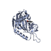
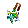
- PDB-5fgp: Crystal structure of D. melanogaster Pur-alpha repeat I-II in com... -
+ Open data

- Basic information
| Entry | Database: PDB / ID: 5fgp | |||||||||
|---|---|---|---|---|---|---|---|---|---|---|
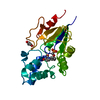
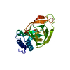
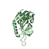
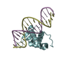
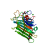
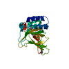
| Title | Crystal structure of D. melanogaster Pur-alpha repeat I-II in complex with DNA. | |||||||||
 Components Components |
| |||||||||
 Keywords Keywords | DNA BINDING PROTEIN / DNA-protein interaction / RNA-protein interaction / DNA unwinding / FXTAS / ALS / FTLD / 5q31.3 microdeletion syndrome / neurodegeneration | |||||||||
| Function / homology |  Function and homology information Function and homology informationpurine-rich negative regulatory element binding / RNA polymerase II transcription regulatory region sequence-specific DNA binding / sequence-specific DNA binding / DNA-binding transcription factor activity, RNA polymerase II-specific / mRNA binding / regulation of transcription by RNA polymerase II / DNA binding / RNA binding / identical protein binding / nucleus / cytoplasm Similarity search - Function | |||||||||
| Biological species |  | |||||||||
| Method |  X-RAY DIFFRACTION / SYNCHROTRON / MOLECULAR REPLACEMENT / Resolution: 2 Å X-RAY DIFFRACTION / SYNCHROTRON / MOLECULAR REPLACEMENT / Resolution: 2 Å | |||||||||
 Authors Authors | Weber, J. / Janowski, R. / Niessing, D. | |||||||||
 Citation Citation | Journal: Elife / Year: 2016 Title: Structural basis of nucleic-acid recognition and double-strand unwinding by the essential neuronal protein Pur-alpha. Authors: Weber, J. / Bao, H. / Hartlmuller, C. / Wang, Z. / Windhager, A. / Janowski, R. / Madl, T. / Jin, P. / Niessing, D. | |||||||||
| History |
|
- Structure visualization
| Structure viewer | Molecule: MolmilJmol/JSmol |
|---|
- Downloads & links
-Download
| PDBx/mmCIF format | 5fgp.cif.gz | 85.2 KB | Display | PDBx/mmCIF format |
|---|---|---|---|---|
| PDB format | pdb5fgp.ent.gz | 62 KB | Display | PDB format |
| PDBx/mmJSON format | 5fgp.json.gz | Tree view | PDBx/mmJSON format | |
| Others |  Other downloads Other downloads |
-Validation report
| Arichive directory | https://data.pdbj.org/pub/pdb/validation_reports/fg/5fgpftp://data.pdbj.org/pub/pdb/validation_reports/fg/5fgp | HTTPS FTP |
|---|
-Related structure data
| Related structure data | 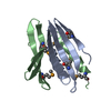 5fgoC 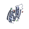 3k44S S: Starting model for refinement C: citing same article ( |
|---|---|
| Similar structure data |


-Links
 PDBj
PDBj- Assembly
| Deposited unit | 
| ||||||||
|---|---|---|---|---|---|---|---|---|---|
| 1 |
| ||||||||
| Unit cell |
| ||||||||
| Components on special symmetry positions |
|
-Components
| #1: Protein | Mass: 17495.152 Da / Num. of mol.: 1 Source method: isolated from a genetically manipulated source Source: (gene. exp.)  |
|---|---|
| #2: DNA chain | Mass: 2179.435 Da / Num. of mol.: 1 / Source method: obtained synthetically / Source: (synth.) |
| #3: Chemical | ChemComp-CL /   Mass: 35.453 Da / Num. of mol.: 1 Mass: 35.453 Da / Num. of mol.: 1Source method: isolated from a genetically manipulated source Formula: Cl / Source: (gene. exp.) |
| #4: Chemical | ChemComp-SO4 /   Mass: 96.063 Da / Num. of mol.: 1 Mass: 96.063 Da / Num. of mol.: 1Source method: isolated from a genetically manipulated source Formula: SO4 / Source: (gene. exp.) |
| #5: Water | ChemComp-HOH /  Mass: 18.015 Da / Num. of mol.: 126 / Source method: isolated from a natural source / Formula: H2O Mass: 18.015 Da / Num. of mol.: 126 / Source method: isolated from a natural source / Formula: H2O |
| Has protein modification | Y |
-Experimental details
-Experiment
| Experiment | Method: X-RAY DIFFRACTION |
|---|
- Sample preparation
| Crystal | Density Matthews: 2.23 Å3/Da / Density % sol: 44.87 % |
|---|---|
| Crystal grow | Temperature: 294 K / Method: vapor diffusion, hanging drop Details: 50 mM MES pH 5.2, 500 mM (NH4)2SO4, 1 mM TCEP and 16 % PEG400 |
-Data collection
| Diffraction | Mean temperature: 100 K |
|---|---|
| Diffraction source | Source: SYNCHROTRON / Site: ESRF  / Beamline: ID23-2 / Wavelength: 0.8726 Å / Beamline: ID23-2 / Wavelength: 0.8726 Å |
| Detector | Type: MARMOSAIC 225 mm CCD / Detector: CCD / Date: May 15, 2011 |
| Radiation | Protocol: SINGLE WAVELENGTH / Monochromatic (M) / Laue (L): M / Scattering type: x-ray |
| Radiation wavelength | Wavelength: 0.8726 Å / Relative weight: 1 |
| Reflection | Resolution: 2→50 Å / Num. obs: 11349 / % possible obs: 99.4 % / Redundancy: 13.1 % / Rmerge(I) obs: 0.125 / Net I/σ(I): 18.85 |
| Reflection shell | Resolution: 2→2.05 Å / Redundancy: 7.6 % / Rmerge(I) obs: 0.793 / Mean I/σ(I) obs: 2.61 / % possible all: 94.3 |
- Processing
| Software |
| |||||||||||||||||||||||||||||||||||||||||||||||||||||||||||||||||||||||||||
|---|---|---|---|---|---|---|---|---|---|---|---|---|---|---|---|---|---|---|---|---|---|---|---|---|---|---|---|---|---|---|---|---|---|---|---|---|---|---|---|---|---|---|---|---|---|---|---|---|---|---|---|---|---|---|---|---|---|---|---|---|---|---|---|---|---|---|---|---|---|---|---|---|---|---|---|---|
| Refinement | Method to determine structure: MOLECULAR REPLACEMENT Starting model: 3K44 Resolution: 2→41.934 Å / SU ML: 0.22 / Cross valid method: FREE R-VALUE / σ(F): 1.36 / Phase error: 21.68 / Stereochemistry target values: ML
| |||||||||||||||||||||||||||||||||||||||||||||||||||||||||||||||||||||||||||
| Solvent computation | Shrinkage radii: 0.9 Å / VDW probe radii: 1.11 Å / Solvent model: FLAT BULK SOLVENT MODEL | |||||||||||||||||||||||||||||||||||||||||||||||||||||||||||||||||||||||||||
| Refinement step | Cycle: LAST / Resolution: 2→41.934 Å
| |||||||||||||||||||||||||||||||||||||||||||||||||||||||||||||||||||||||||||
| Refine LS restraints |
| |||||||||||||||||||||||||||||||||||||||||||||||||||||||||||||||||||||||||||
| LS refinement shell |
| |||||||||||||||||||||||||||||||||||||||||||||||||||||||||||||||||||||||||||
| Refinement TLS params. | Method: refined / Refine-ID: X-RAY DIFFRACTION
| |||||||||||||||||||||||||||||||||||||||||||||||||||||||||||||||||||||||||||
| Refinement TLS group |
|
