Mass: 18.015 Da / Num. of mol.: 180 / Source method: isolated from a natural source / Formula: H2O
Has protein modification
Y
-
Experimental details
-
Experiment
Experiment
Method: X-RAY DIFFRACTION / Number of used crystals: 1
-
Sample preparation
Crystal
Density Matthews: 2.19 Å3/Da / Density % sol: 43.86 %
Crystal grow
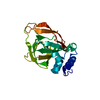
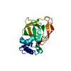
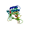
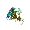
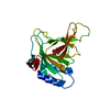
Temperature: 297 K / Method: vapor diffusion, hanging drop / pH: 5.6 Details: Hanging drops of 1.5 microL of approximately 8 mg/mL CDO and 1.5 microL reservoir buffer were allowed to equilibrate above the reservoir buffer (24-34% (w/v) polyethylene glycol 4000, 100- ...Details: Hanging drops of 1.5 microL of approximately 8 mg/mL CDO and 1.5 microL reservoir buffer were allowed to equilibrate above the reservoir buffer (24-34% (w/v) polyethylene glycol 4000, 100-250 mM ammonium acetate, 100 mM sodium citrate, 0-4 mM dithionite, 40 mM 3-mercaptopropionic acid). Crystals grew as needles or starbursts of approximately 0.1 mm in length in one week, pH 5.6, VAPOR DIFFUSION, HANGING DROP, temperature 297K
-
Data collection
Diffraction
Mean temperature: 100 K
Diffraction source
Source: SYNCHROTRON / Site: Australian Synchrotron / Beamline: MX1 / Wavelength: 0.9537 Å
Detector
Type: ADSC QUANTUM 210r / Detector: CCD / Date: Mar 31, 2012 / Details: Double Si with sagittaly bent second crystal
Radiation
Monochromator: Silicon Double Crystal / Protocol: SINGLE WAVELENGTH / Monochromatic (M) / Laue (L): M / Scattering type: x-ray
In the structure databanks used in Yorodumi, some data are registered as the other names, "COVID-19 virus" and "2019-nCoV". Here are the details of the virus and the list of structure data.
Jan 31, 2019. EMDB accession codes are about to change! (news from PDBe EMDB page)
EMDB accession codes are about to change! (news from PDBe EMDB page)
The allocation of 4 digits for EMDB accession codes will soon come to an end. Whilst these codes will remain in use, new EMDB accession codes will include an additional digit and will expand incrementally as the available range of codes is exhausted. The current 4-digit format prefixed with “EMD-” (i.e. EMD-XXXX) will advance to a 5-digit format (i.e. EMD-XXXXX), and so on. It is currently estimated that the 4-digit codes will be depleted around Spring 2019, at which point the 5-digit format will come into force.
The EM Navigator/Yorodumi systems omit the EMD- prefix.
Related info.:Q: What is EMD? / ID/Accession-code notation in Yorodumi/EM Navigator
Yorodumi is a browser for structure data from EMDB, PDB, SASBDB, etc.
This page is also the successor to EM Navigator detail page, and also detail information page/front-end page for Omokage search.
The word "yorodu" (or yorozu) is an old Japanese word meaning "ten thousand". "mi" (miru) is to see.
Related info.:EMDB / PDB / SASBDB / Comparison of 3 databanks / Yorodumi Search / Aug 31, 2016. New EM Navigator & Yorodumi / Yorodumi Papers / Jmol/JSmol / Function and homology information / Changes in new EM Navigator and Yorodumi
 Movie
Movie Controller

 Yorodumi
 Open data
 Basic information
 Components
 Keywords
 Function and homology information
 X-RAY DIFFRACTION /
  Authors
 Citation
 Structure visualization
 Downloads & links
 Other downloads


 PDBj
PDBj


 Assembly
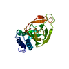


 Mass: 55.845 Da / Num. of mol.: 1 / Source method: obtained synthetically / Formula: Fe
 Mass: 138.209 Da / Num. of mol.: 1 / Source method: obtained synthetically / Formula: C3H6O2S2
 Mass: 18.015 Da / Num. of mol.: 180 / Source method: isolated from a natural source / Formula: H2O
 Sample preparation
 / Beamline: MX1 / Wavelength: 0.9537 Å
 Processing