 Movie
Movie Controller

[English] 日本語
 Yorodumi
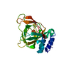
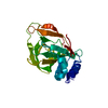
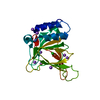
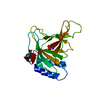
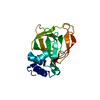
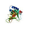
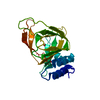
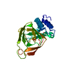
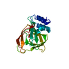
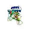
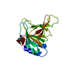
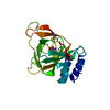
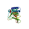
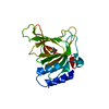
- PDB-4ies: Cys-persulfenate bound Cysteine Dioxygenase at pH 6.2 in the pres... -
+ Open data

- Basic information
| Entry | Database: PDB / ID: 4ies | ||||||
|---|---|---|---|---|---|---|---|
| Title | Cys-persulfenate bound Cysteine Dioxygenase at pH 6.2 in the presence of Cys | ||||||
 Components Components | Cysteine dioxygenase type 1 | ||||||
 Keywords Keywords | OXIDOREDUCTASE / Cupin fold / catalyzes oxidation / cysteine to cysteine sulfinate / C93-Y157 crosslink / Cytosol | ||||||
| Function / homology |  Function and homology information Function and homology information: / Degradation of cysteine and homocysteine / taurine biosynthetic process / cysteine dioxygenase / cysteine dioxygenase activity / response to azide / L-cysteine catabolic process / : / response to glucagon / nickel cation binding ...: / Degradation of cysteine and homocysteine / taurine biosynthetic process / cysteine dioxygenase / cysteine dioxygenase activity / response to azide / L-cysteine catabolic process / : / response to glucagon / nickel cation binding / response to amino acid / response to cAMP / lactation / response to glucocorticoid / ferrous iron binding / response to ethanol / zinc ion binding / cytosol Similarity search - Function | ||||||
| Biological species |  | ||||||
| Method |  X-RAY DIFFRACTION / SYNCHROTRON / MOLECULAR REPLACEMENT / Resolution: 1.4 Å X-RAY DIFFRACTION / SYNCHROTRON / MOLECULAR REPLACEMENT / Resolution: 1.4 Å | ||||||
 Authors Authors | Driggers, C.M. / Cooley, R.B. / Sankaran, B. / Karplus, P.A. | ||||||
 Citation Citation | Journal: J.Mol.Biol. / Year: 2013 Title: Cysteine Dioxygenase Structures from pH4 to 9: Consistent Cys-Persulfenate Formation at Intermediate pH and a Cys-Bound Enzyme at Higher pH. Authors: Driggers, C.M. / Cooley, R.B. / Sankaran, B. / Hirschberger, L.L. / Stipanuk, M.H. / Karplus, P.A. | ||||||
| History |
|
- Structure visualization
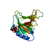
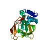
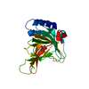
| Structure viewer | Molecule: MolmilJmol/JSmol |
|---|
- Downloads & links
-Download
| PDBx/mmCIF format | 4ies.cif.gz | 103.4 KB | Display | PDBx/mmCIF format |
|---|---|---|---|---|
| PDB format | pdb4ies.ent.gz | 79.5 KB | Display | PDB format |
| PDBx/mmJSON format | 4ies.json.gz | Tree view | PDBx/mmJSON format | |
| Others |  Other downloads Other downloads |
-Validation report
| Arichive directory | https://data.pdbj.org/pub/pdb/validation_reports/ie/4iesftp://data.pdbj.org/pub/pdb/validation_reports/ie/4ies | HTTPS FTP |
|---|
-Related structure data
| Related structure data |  4ieoC 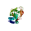 4iepC 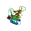 4ieqC 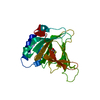 4ierC  4ietC  4ieuC  4ievC  4iewC 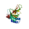 4iexC  4ieyC  4iezC  4jtnC  4jtoC  4if0 4if1 C: citing same article ( |
|---|---|
| Similar structure data |


-Links
 PDBj
PDBj- Assembly
| Deposited unit | 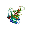
| ||||||||
|---|---|---|---|---|---|---|---|---|---|
| 1 |
| ||||||||
| Unit cell |
| ||||||||
| Components on special symmetry positions |
|
-Components
| #1: Protein | Mass: 23058.889 Da / Num. of mol.: 1 Source method: isolated from a genetically manipulated source Source: (gene. exp.)  |
|---|---|
| #2: Chemical | ChemComp-FE /   Mass: 55.845 Da / Num. of mol.: 1 / Source method: obtained synthetically / Formula: Fe Mass: 55.845 Da / Num. of mol.: 1 / Source method: obtained synthetically / Formula: Fe |
| #3: Chemical | ChemComp-2CO / 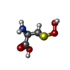  Type: L-peptide linking / Mass: 153.157 Da / Num. of mol.: 1 / Source method: obtained synthetically / Formula: C3H7NO4S Type: L-peptide linking / Mass: 153.157 Da / Num. of mol.: 1 / Source method: obtained synthetically / Formula: C3H7NO4S |
| #4: Water | ChemComp-HOH /  Mass: 18.015 Da / Num. of mol.: 240 / Source method: isolated from a natural source / Formula: H2O Mass: 18.015 Da / Num. of mol.: 240 / Source method: isolated from a natural source / Formula: H2O |
| Has protein modification | Y |
-Experimental details
-Experiment
| Experiment | Method: X-RAY DIFFRACTION / Number of used crystals: 1 |
|---|
- Sample preparation
| Crystal | Density Matthews: 2.2 Å3/Da / Density % sol: 44.13 % |
|---|---|
| Crystal grow | Temperature: 298 K / Method: vapor diffusion, hanging drop / pH: 6.2 Details: Purified enzyme was concentrated to ~8 mg/mL and then added into a crystallization screen containing 0.1 M tri-sodium citrate pH=5.6, 24-34% PEG 4K, and 0.1-0.25 M ammonium acetate. 1.5L of ...Details: Purified enzyme was concentrated to ~8 mg/mL and then added into a crystallization screen containing 0.1 M tri-sodium citrate pH=5.6, 24-34% PEG 4K, and 0.1-0.25 M ammonium acetate. 1.5L of protein solution was added to each well and mixed with an equivalent volume of reservoir solution., pH 6.2, VAPOR DIFFUSION, HANGING DROP, temperature 298K |
-Data collection
| Diffraction | Mean temperature: 100 K |
|---|---|
| Diffraction source | Source: SYNCHROTRON / Site: ALS  / Beamline: 5.0.1 / Wavelength: 0.976 Å / Beamline: 5.0.1 / Wavelength: 0.976 Å |
| Detector | Type: ADSC QUANTUM 315r / Detector: CCD / Date: Dec 4, 2010 |
| Radiation | Monochromator: Si(220) Asymmetric cut single crystal / Protocol: SINGLE WAVELENGTH / Monochromatic (M) / Laue (L): M / Scattering type: x-ray |
| Radiation wavelength | Wavelength: 0.976 Å / Relative weight: 1 |
| Reflection | Resolution: 1.4→29 Å / Num. obs: 41131 / % possible obs: 99.1 % / Observed criterion σ(F): 0 / Observed criterion σ(I): 0 |
- Processing
| Software |
| ||||||||||||||||||||||||||||||||||||||||||||||||||||||||||||||||||||||||||||||||||||||||||||||||||||||||||||||||||||||||||||||||||||||||||||||||||||||||||||||||||||||||||||||||||||||||||||||||||||||||||||||||||
|---|---|---|---|---|---|---|---|---|---|---|---|---|---|---|---|---|---|---|---|---|---|---|---|---|---|---|---|---|---|---|---|---|---|---|---|---|---|---|---|---|---|---|---|---|---|---|---|---|---|---|---|---|---|---|---|---|---|---|---|---|---|---|---|---|---|---|---|---|---|---|---|---|---|---|---|---|---|---|---|---|---|---|---|---|---|---|---|---|---|---|---|---|---|---|---|---|---|---|---|---|---|---|---|---|---|---|---|---|---|---|---|---|---|---|---|---|---|---|---|---|---|---|---|---|---|---|---|---|---|---|---|---|---|---|---|---|---|---|---|---|---|---|---|---|---|---|---|---|---|---|---|---|---|---|---|---|---|---|---|---|---|---|---|---|---|---|---|---|---|---|---|---|---|---|---|---|---|---|---|---|---|---|---|---|---|---|---|---|---|---|---|---|---|---|---|---|---|---|---|---|---|---|---|---|---|---|---|---|---|---|---|
| Refinement | Method to determine structure: MOLECULAR REPLACEMENT / Resolution: 1.4→28.034 Å / SU ML: 0.19 / σ(F): 1.33 / Phase error: 20.08 / Stereochemistry target values: ML
| ||||||||||||||||||||||||||||||||||||||||||||||||||||||||||||||||||||||||||||||||||||||||||||||||||||||||||||||||||||||||||||||||||||||||||||||||||||||||||||||||||||||||||||||||||||||||||||||||||||||||||||||||||
| Solvent computation | Shrinkage radii: 0.9 Å / VDW probe radii: 1.11 Å / Solvent model: FLAT BULK SOLVENT MODEL | ||||||||||||||||||||||||||||||||||||||||||||||||||||||||||||||||||||||||||||||||||||||||||||||||||||||||||||||||||||||||||||||||||||||||||||||||||||||||||||||||||||||||||||||||||||||||||||||||||||||||||||||||||
| Refinement step | Cycle: LAST / Resolution: 1.4→28.034 Å
| ||||||||||||||||||||||||||||||||||||||||||||||||||||||||||||||||||||||||||||||||||||||||||||||||||||||||||||||||||||||||||||||||||||||||||||||||||||||||||||||||||||||||||||||||||||||||||||||||||||||||||||||||||
| Refine LS restraints |
| ||||||||||||||||||||||||||||||||||||||||||||||||||||||||||||||||||||||||||||||||||||||||||||||||||||||||||||||||||||||||||||||||||||||||||||||||||||||||||||||||||||||||||||||||||||||||||||||||||||||||||||||||||
| LS refinement shell |
|
