 Movie
Movie Controller


+ Open data

- Basic information
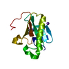
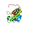
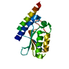
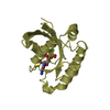
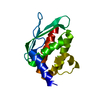
| Entry | Database: PDB / ID: 5fgo | ||||||
|---|---|---|---|---|---|---|---|
| Title | Crystal structure of D. melanogaster Pur-alpha repeat III. | ||||||
 Components Components | CG1507-PB, isoform B | ||||||
 Keywords Keywords | DNA BINDING PROTEIN / DNA-protein interaction / RNA-protein interaction / DNA unwinding / FXTAS / ALS / FTLD / 5q31.3 microdeletion syndrome / neurodegeneration | ||||||
| Function / homology |  Function and homology information Function and homology informationpurine-rich negative regulatory element binding / RNA polymerase II transcription regulatory region sequence-specific DNA binding / sequence-specific DNA binding / DNA-binding transcription factor activity, RNA polymerase II-specific / mRNA binding / regulation of transcription by RNA polymerase II / DNA binding / RNA binding / identical protein binding / nucleus / cytoplasm Similarity search - Function | ||||||
| Biological species |  | ||||||
| Method |  X-RAY DIFFRACTION / SYNCHROTRON / MOLECULAR REPLACEMENT / Resolution: 2.6 Å X-RAY DIFFRACTION / SYNCHROTRON / MOLECULAR REPLACEMENT / Resolution: 2.6 Å | ||||||
 Authors Authors | Windhager, A. / Janowski, R. / Niessing, D. | ||||||
 Citation Citation | Journal: Elife / Year: 2016 Title: Structural basis of nucleic-acid recognition and double-strand unwinding by the essential neuronal protein Pur-alpha. Authors: Weber, J. / Bao, H. / Hartlmuller, C. / Wang, Z. / Windhager, A. / Janowski, R. / Madl, T. / Jin, P. / Niessing, D. | ||||||
| History |
|
- Structure visualization
| Structure viewer | Molecule: MolmilJmol/JSmol |
|---|
- Downloads & links
-Download
| PDBx/mmCIF format | 5fgo.cif.gz | 99.6 KB | Display | PDBx/mmCIF format |
|---|---|---|---|---|
| PDB format | pdb5fgo.ent.gz | 77.1 KB | Display | PDB format |
| PDBx/mmJSON format | 5fgo.json.gz | Tree view | PDBx/mmJSON format | |
| Others |  Other downloads Other downloads |
-Validation report
| Arichive directory | https://data.pdbj.org/pub/pdb/validation_reports/fg/5fgoftp://data.pdbj.org/pub/pdb/validation_reports/fg/5fgo | HTTPS FTP |
|---|
-Related structure data
| Related structure data | 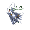 5fgpC 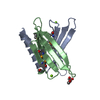 3n8bS S: Starting model for refinement C: citing same article ( |
|---|---|
| Similar structure data |


-Links
 PDBj
PDBj- Assembly
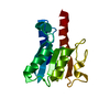
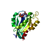
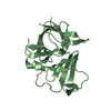
| Deposited unit | 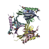
| ||||||||
|---|---|---|---|---|---|---|---|---|---|
| 1 | 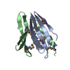
| ||||||||
| 2 | 
| ||||||||
| 3 | 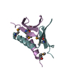
| ||||||||
| Unit cell |
|
-Components
| #1: Protein | Mass: 9698.516 Da / Num. of mol.: 6 Source method: isolated from a genetically manipulated source Source: (gene. exp.)  #2: Chemical |   Mass: 35.453 Da / Num. of mol.: 2 / Source method: obtained synthetically / Formula: Cl Mass: 35.453 Da / Num. of mol.: 2 / Source method: obtained synthetically / Formula: Cl#3: Water | ChemComp-HOH / |  Mass: 18.015 Da / Num. of mol.: 170 / Source method: isolated from a natural source / Formula: H2O Mass: 18.015 Da / Num. of mol.: 170 / Source method: isolated from a natural source / Formula: H2OHas protein modification | Y | |
|---|
-Experimental details
-Experiment
| Experiment | Method: X-RAY DIFFRACTION |
|---|
- Sample preparation
| Crystal | Density Matthews: 1.98 Å3/Da / Density % sol: 37.87 % |
|---|---|
| Crystal grow | Temperature: 294 K / Method: vapor diffusion, hanging drop Details: 50 mM MES pH 6.5, 200 mM NaCl, 16% PEG 3350 and 6 % MPD |
-Data collection
| Diffraction | Mean temperature: 100 K |
|---|---|
| Diffraction source | Source: SYNCHROTRON / Site: ESRF  / Beamline: ID14-1 / Wavelength: 0.9334 Å / Beamline: ID14-1 / Wavelength: 0.9334 Å |
| Detector | Type: ADSC QUANTUM 210 / Detector: CCD / Date: Jul 9, 2013 |
| Radiation | Protocol: MAD / Monochromatic (M) / Laue (L): M / Scattering type: x-ray |
| Radiation wavelength | Wavelength: 0.9334 Å / Relative weight: 1 |
| Reflection | Resolution: 2.6→50 Å / Num. obs: 13999 / % possible obs: 96.5 % / Redundancy: 1.9 % / Rmerge(I) obs: 0.11 / Net I/σ(I): 19.4 |
| Reflection shell | Resolution: 2.6→2.65 Å / Redundancy: 1.9 % / Rmerge(I) obs: 0.68 / Mean I/σ(I) obs: 1.97 / % possible all: 98.5 |
- Processing
| Software |
| ||||||||||||||||||||||||||||||||||||||||||||||||||||||||||||||||||||||
|---|---|---|---|---|---|---|---|---|---|---|---|---|---|---|---|---|---|---|---|---|---|---|---|---|---|---|---|---|---|---|---|---|---|---|---|---|---|---|---|---|---|---|---|---|---|---|---|---|---|---|---|---|---|---|---|---|---|---|---|---|---|---|---|---|---|---|---|---|---|---|---|
| Refinement | Method to determine structure: MOLECULAR REPLACEMENT Starting model: 3N8B Resolution: 2.6→47.725 Å / SU ML: 0.37 / Cross valid method: FREE R-VALUE / σ(F): 1.12 / Phase error: 28.3 / Stereochemistry target values: ML
| ||||||||||||||||||||||||||||||||||||||||||||||||||||||||||||||||||||||
| Solvent computation | Shrinkage radii: 0.9 Å / VDW probe radii: 1.11 Å / Solvent model: FLAT BULK SOLVENT MODEL | ||||||||||||||||||||||||||||||||||||||||||||||||||||||||||||||||||||||
| Refinement step | Cycle: LAST / Resolution: 2.6→47.725 Å
| ||||||||||||||||||||||||||||||||||||||||||||||||||||||||||||||||||||||
| Refine LS restraints |
| ||||||||||||||||||||||||||||||||||||||||||||||||||||||||||||||||||||||
| LS refinement shell |
|
