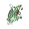
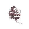
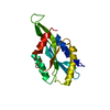
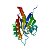
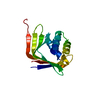
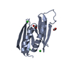
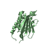
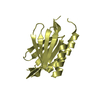
- PDB-3k44: Crystal Structure of Drosophila melanogaster Pur-alpha -
+
Open data
ID or keywords:
Loading...
-
Basic information
Entry
Database: PDB / ID: 3k44
Title
Crystal Structure of Drosophila melanogaster Pur-alpha
Components
Purine-rich binding protein-alpha, isoform B
Keywords
NUCLEIC ACID BINDING PROTEIN / Pur-alpha / Pur repeat / Pur domain / Whirly fold / DNA BINDING PROTEIN / RNA BINDING PROTEIN
Function / homology
Function and homology information
purine-rich negative regulatory element binding / RNA polymerase II transcription regulatory region sequence-specific DNA binding / sequence-specific DNA binding / DNA-binding transcription factor activity, RNA polymerase II-specific / mRNA binding / regulation of transcription by RNA polymerase II / DNA binding / RNA binding / identical protein binding / nucleus / cytoplasm Similarity search - Function
Secreted effector protein pipB2 fold - #30 / Secreted effector protein pipB2 fold / PurA ssDNA and RNA-binding protein / Purine-rich element binding protein family / DNA/RNA-binding repeats in PUR-alpha/beta/gamma and in hypothetical proteins from spirochetes and the Bacteroides-Cytophaga-Flexibacter bacteria. / 2-Layer Sandwich / Alpha Beta Similarity search - Domain/homology
In the structure databanks used in Yorodumi, some data are registered as the other names, "COVID-19 virus" and "2019-nCoV". Here are the details of the virus and the list of structure data.
Jan 31, 2019. EMDB accession codes are about to change! (news from PDBe EMDB page)
EMDB accession codes are about to change! (news from PDBe EMDB page)
The allocation of 4 digits for EMDB accession codes will soon come to an end. Whilst these codes will remain in use, new EMDB accession codes will include an additional digit and will expand incrementally as the available range of codes is exhausted. The current 4-digit format prefixed with “EMD-” (i.e. EMD-XXXX) will advance to a 5-digit format (i.e. EMD-XXXXX), and so on. It is currently estimated that the 4-digit codes will be depleted around Spring 2019, at which point the 5-digit format will come into force.
The EM Navigator/Yorodumi systems omit the EMD- prefix.
Related info.:Q: What is EMD? / ID/Accession-code notation in Yorodumi/EM Navigator
Yorodumi is a browser for structure data from EMDB, PDB, SASBDB, etc.
This page is also the successor to EM Navigator detail page, and also detail information page/front-end page for Omokage search.
The word "yorodu" (or yorozu) is an old Japanese word meaning "ten thousand". "mi" (miru) is to see.
Related info.:EMDB / PDB / SASBDB / Comparison of 3 databanks / Yorodumi Search / Aug 31, 2016. New EM Navigator & Yorodumi / Yorodumi Papers / Jmol/JSmol / Function and homology information / Changes in new EM Navigator and Yorodumi
 Movie
Movie Controller


 Open data
 Basic information
 Components
 Keywords
 Function and homology information
 X-RAY DIFFRACTION /
  Authors
 Citation
 Structure visualization
 Downloads & links
 Other downloads
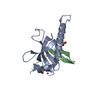

 PDBj
PDBj Assembly
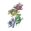


 Mass: 35.453 Da / Num. of mol.: 8 / Source method: obtained synthetically / Formula: Cl
 Mass: 62.068 Da / Num. of mol.: 4 / Source method: obtained synthetically / Formula: C2H6O2
 Mass: 18.015 Da / Num. of mol.: 156 / Source method: isolated from a natural source / Formula: H2O
 Sample preparation
 Processing