 Movie
Movie Controller

[English] 日本語
 Yorodumi
- PDB-1f47: THE BACTERIAL CELL-DIVISION PROTEIN ZIPA AND ITS INTERACTION WITH... -
+ Open data

- Basic information
| Entry | Database: PDB / ID: 1f47 | ||||||
|---|---|---|---|---|---|---|---|
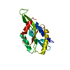
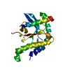
| Title | THE BACTERIAL CELL-DIVISION PROTEIN ZIPA AND ITS INTERACTION WITH AN FTSZ FRAGMENT REVEALED BY X-RAY CRYSTALLOGRAPHY | ||||||
 Components Components |
| ||||||
 Keywords Keywords | CELL CYCLE / cell division zipa / CELL DIVISiON ftsz / SEPARATION / INNER MEMBRANE / TRANSMEMBRANE | ||||||
| Function / homology |  Function and homology information Function and homology informationdivisome complex / division septum assembly / FtsZ-dependent cytokinesis / cell division site / protein polymerization / cell division / GTPase activity / GTP binding / protein homodimerization activity / identical protein binding ...divisome complex / division septum assembly / FtsZ-dependent cytokinesis / cell division site / protein polymerization / cell division / GTPase activity / GTP binding / protein homodimerization activity / identical protein binding / plasma membrane / cytoplasm Similarity search - Function | ||||||
| Biological species |  | ||||||
| Method |  X-RAY DIFFRACTION / Resolution: 1.95 Å X-RAY DIFFRACTION / Resolution: 1.95 Å | ||||||
 Authors Authors | Mosyak, L. / Zhang, Y. / Glasfeld, E. / Stahl, M. / Somers, W.S. | ||||||
 Citation Citation | Journal: EMBO J. / Year: 2000 Title: The bacterial cell-division protein ZipA and its interaction with an FtsZ fragment revealed by X-ray crystallography. Authors: Mosyak, L. / Zhang, Y. / Glasfeld, E. / Haney, S. / Stahl, M. / Seehra, J. / Somers, W.S. | ||||||
| History |
|
- Structure visualization
| Structure viewer | Molecule: MolmilJmol/JSmol |
|---|
- Downloads & links
-Download
| PDBx/mmCIF format | 1f47.cif.gz | 47.5 KB | Display | PDBx/mmCIF format |
|---|---|---|---|---|
| PDB format | pdb1f47.ent.gz | 33.8 KB | Display | PDB format |
| PDBx/mmJSON format | 1f47.json.gz | Tree view | PDBx/mmJSON format | |
| Others |  Other downloads Other downloads |
-Validation report
| Arichive directory | https://data.pdbj.org/pub/pdb/validation_reports/f4/1f47ftp://data.pdbj.org/pub/pdb/validation_reports/f4/1f47 | HTTPS FTP |
|---|
-Related structure data


-Links
 PDBj
PDBj


- Assembly
| Deposited unit | 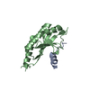
| ||||||||
|---|---|---|---|---|---|---|---|---|---|
| 1 |
| ||||||||
| Unit cell |
|
-Components
| #1: Protein | Mass: 16134.484 Da / Num. of mol.: 1 Source method: isolated from a genetically manipulated source Source: (gene. exp.) |
|---|---|
| #2: Protein/peptide | Mass: 2022.281 Da / Num. of mol.: 1 Source method: isolated from a genetically manipulated source Source: (gene. exp.) |
| #3: Water | ChemComp-HOH /  Mass: 18.015 Da / Num. of mol.: 203 / Source method: isolated from a natural source / Formula: H2O Mass: 18.015 Da / Num. of mol.: 203 / Source method: isolated from a natural source / Formula: H2O |
-Experimental details
-Experiment
| Experiment | Method: X-RAY DIFFRACTION / Number of used crystals: 1 |
|---|
- Sample preparation
| Crystal | Density Matthews: 2.07 Å3/Da / Density % sol: 40.57 % | ||||||||||||||||||||
|---|---|---|---|---|---|---|---|---|---|---|---|---|---|---|---|---|---|---|---|---|---|
| Crystal grow | Temperature: 291 K / Method: vapor diffusion, hanging drop / pH: 9 Details: 30% PEG 6000, 100 mM Bicine, pH 9.0, VAPOR DIFFUSION, HANGING DROP, temperature 18K | ||||||||||||||||||||
| Crystal grow | *PLUS pH: 6 | ||||||||||||||||||||
| Components of the solutions | *PLUS
|
-Data collection
| Diffraction | Mean temperature: 100 K |
|---|---|
| Diffraction source | Source: ROTATING ANODE / Type: RIGAKU RU200 / Wavelength: 1.5418 |
| Detector | Type: RIGAKU RAXIS / Detector: IMAGE PLATE / Date: Dec 12, 1999 |
| Radiation | Protocol: SINGLE WAVELENGTH / Monochromatic (M) / Laue (L): M / Scattering type: x-ray |
| Radiation wavelength | Wavelength: 1.5418 Å / Relative weight: 1 |
| Reflection | Resolution: 1.95→25 Å / Num. all: 10556 / Num. obs: 10556 / % possible obs: 95.6 % / Observed criterion σ(F): 0 / Observed criterion σ(I): 0 / Redundancy: 3.92 % / Biso Wilson estimate: 30 Å2 / Rmerge(I) obs: 0.055 / Net I/σ(I): 26.9 |
| Reflection shell | Resolution: 1.95→2.02 Å / Redundancy: 2.5 % / Rmerge(I) obs: 0.146 / % possible all: 88.6 |
| Reflection | *PLUS |
| Reflection shell | *PLUS % possible obs: 88.6 % / Mean I/σ(I) obs: 8.6 |
- Processing
| Software |
| ||||||||||||||||||||
|---|---|---|---|---|---|---|---|---|---|---|---|---|---|---|---|---|---|---|---|---|---|
| Refinement | Resolution: 1.95→25 Å / σ(F): 0 / σ(I): 0 / Stereochemistry target values: Engh & Huber
| ||||||||||||||||||||
| Refinement step | Cycle: LAST / Resolution: 1.95→25 Å
| ||||||||||||||||||||
| Refine LS restraints |
| ||||||||||||||||||||
| Software | *PLUS Name: CNS / Classification: refinement | ||||||||||||||||||||
| Refinement | *PLUS Lowest resolution: 25 Å / σ(F): 0 / % reflection Rfree: 5 % | ||||||||||||||||||||
| Solvent computation | *PLUS | ||||||||||||||||||||
| Displacement parameters | *PLUS |
