 Movie
Movie Controller


+ Open data

- Basic information
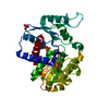
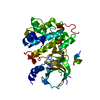
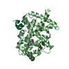
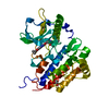
| Entry | Database: PDB / ID: 5v3n | ||||||
|---|---|---|---|---|---|---|---|
| Title | Structure of S. cerevisiae Ulp2-Tof2-Csm1 complex | ||||||
 Components Components |
| ||||||
 Keywords Keywords | HYDROLASE / monopolin / cohibin / rDNA silencing / desumoylation | ||||||
| Function / homology |  Function and homology information Function and homology informationubiquitin-like protein peptidase activity / microtubule site clamp / monopolin complex / chromosome, centromeric core domain / meiotic sister chromatid segregation / homologous chromosome segregation / protein localization to nucleolar rDNA repeats / spindle attachment to meiosis I kinetochore / meiotic chromosome segregation / rDNA chromatin condensation ...ubiquitin-like protein peptidase activity / microtubule site clamp / monopolin complex / chromosome, centromeric core domain / meiotic sister chromatid segregation / homologous chromosome segregation / protein localization to nucleolar rDNA repeats / spindle attachment to meiosis I kinetochore / meiotic chromosome segregation / rDNA chromatin condensation / meiotic sister chromatid cohesion, centromeric / phosphatase activator activity / rDNA binding / attachment of mitotic spindle microtubules to kinetochore / rDNA heterochromatin formation / nucleolus organization / cysteine-type peptidase activity / mitotic spindle / nuclear envelope / nucleolus / mitochondrion / proteolysis / identical protein binding Similarity search - Function | ||||||
| Biological species |  | ||||||
| Method |  X-RAY DIFFRACTION / SYNCHROTRON / MOLECULAR REPLACEMENT / Resolution: 1.3 Å X-RAY DIFFRACTION / SYNCHROTRON / MOLECULAR REPLACEMENT / Resolution: 1.3 Å | ||||||
 Authors Authors | SIngh, N. / Corbett, K.D. | ||||||
| Funding support |  United States, 1items United States, 1items
| ||||||
 Citation Citation | Journal: Genes Dev. / Year: 2017 Title: Recruitment of a SUMO isopeptidase to rDNA stabilizes silencing complexes by opposing SUMO targeted ubiquitin ligase activity. Authors: Liang, J. / Singh, N. / Carlson, C.R. / Albuquerque, C.P. / Corbett, K.D. / Zhou, H. | ||||||
| History |
|
- Structure visualization
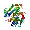
| Structure viewer | Molecule: MolmilJmol/JSmol |
|---|
- Downloads & links
-Download
| PDBx/mmCIF format | 5v3n.cif.gz | 99.2 KB | Display | PDBx/mmCIF format |
|---|---|---|---|---|
| PDB format | pdb5v3n.ent.gz | 75.5 KB | Display | PDB format |
| PDBx/mmJSON format | 5v3n.json.gz | Tree view | PDBx/mmJSON format | |
| Others |  Other downloads Other downloads |
-Validation report
| Arichive directory | https://data.pdbj.org/pub/pdb/validation_reports/v3/5v3nftp://data.pdbj.org/pub/pdb/validation_reports/v3/5v3n | HTTPS FTP |
|---|
-Related structure data
| Related structure data |  5v1aC 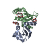 3n4sS S: Starting model for refinement C: citing same article ( |
|---|---|
| Similar structure data |


-Links
 PDBj
PDBj- Assembly
| Deposited unit | 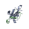
| |||||||||
|---|---|---|---|---|---|---|---|---|---|---|
| 1 | 
| |||||||||
| Unit cell |
| |||||||||
| Components on special symmetry positions |
|
-Components
| #1: Protein | Mass: 12991.447 Da / Num. of mol.: 1 Source method: isolated from a genetically manipulated source Source: (gene. exp.) Strain: ATCC 204508 / S288c / Gene: CSM1, SPO86, YCR086W, YCR86W / Production host:  |
|---|---|
| #2: Protein/peptide | Mass: 4269.733 Da / Num. of mol.: 1 Source method: isolated from a genetically manipulated source Source: (gene. exp.) Strain: VIN7, ATCC 204508 / S288c / Gene: VIN7_2425, TOF2, YKR010C, YK109 / Production host: |
| #3: Water | ChemComp-HOH /  Mass: 18.015 Da / Num. of mol.: 85 / Source method: isolated from a natural source / Formula: H2O Mass: 18.015 Da / Num. of mol.: 85 / Source method: isolated from a natural source / Formula: H2O |
-Experimental details
-Experiment
| Experiment | Method: X-RAY DIFFRACTION / Number of used crystals: 1 |
|---|
- Sample preparation
| Crystal | Density Matthews: 2.09 Å3/Da / Density % sol: 41.04 % |
|---|---|
| Crystal grow | Temperature: 293 K / Method: vapor diffusion, hanging drop Details: 0.2 M Ammonium Acetate, 0.1 M HEPES pH 7.5 and 25% PEG 3350 and 25% Glycerol |
-Data collection
| Diffraction | Mean temperature: 100 K |
|---|---|
| Diffraction source | Source: SYNCHROTRON / Site: APS / Beamline: 24-ID-C / Wavelength: 0.9791 Å |
| Detector | Type: DECTRIS PILATUS3 S 6M / Detector: PIXEL / Date: Nov 28, 2016 |
| Radiation | Protocol: SINGLE WAVELENGTH / Monochromatic (M) / Laue (L): M / Scattering type: x-ray |
| Radiation wavelength | Wavelength: 0.9791 Å / Relative weight: 1 |
| Reflection | Resolution: 1.3→42 Å / Num. obs: 33373 / % possible obs: 96.4 % / Redundancy: 3.7 % / CC1/2: 0.995 / Rmerge(I) obs: 0.068 / Rpim(I) all: 0.042 / Net I/σ(I): 10.1 |
| Reflection shell | Resolution: 1.3→1.32 Å / Redundancy: 3.5 % / Rmerge(I) obs: 2.281 / Mean I/σ(I) obs: 1.1 / CC1/2: 0.418 / Rpim(I) all: 1.418 / % possible all: 92.7 |
- Processing
| Software |
| |||||||||||||||||||||||||||||||||||||||||||||||||||||||||||||||||||||||||||||||||||||||||||||||||||||||||||||||||||||||||||||||||||||||||||||||||||||||||||||||||
|---|---|---|---|---|---|---|---|---|---|---|---|---|---|---|---|---|---|---|---|---|---|---|---|---|---|---|---|---|---|---|---|---|---|---|---|---|---|---|---|---|---|---|---|---|---|---|---|---|---|---|---|---|---|---|---|---|---|---|---|---|---|---|---|---|---|---|---|---|---|---|---|---|---|---|---|---|---|---|---|---|---|---|---|---|---|---|---|---|---|---|---|---|---|---|---|---|---|---|---|---|---|---|---|---|---|---|---|---|---|---|---|---|---|---|---|---|---|---|---|---|---|---|---|---|---|---|---|---|---|---|---|---|---|---|---|---|---|---|---|---|---|---|---|---|---|---|---|---|---|---|---|---|---|---|---|---|---|---|---|---|---|---|
| Refinement | Method to determine structure: MOLECULAR REPLACEMENT Starting model: 3N4S Resolution: 1.3→35.115 Å / SU ML: 0.19 / Cross valid method: FREE R-VALUE / σ(F): 0.03 / Phase error: 32.73
| |||||||||||||||||||||||||||||||||||||||||||||||||||||||||||||||||||||||||||||||||||||||||||||||||||||||||||||||||||||||||||||||||||||||||||||||||||||||||||||||||
| Solvent computation | Shrinkage radii: 0.9 Å / VDW probe radii: 1.11 Å | |||||||||||||||||||||||||||||||||||||||||||||||||||||||||||||||||||||||||||||||||||||||||||||||||||||||||||||||||||||||||||||||||||||||||||||||||||||||||||||||||
| Refinement step | Cycle: LAST / Resolution: 1.3→35.115 Å
| |||||||||||||||||||||||||||||||||||||||||||||||||||||||||||||||||||||||||||||||||||||||||||||||||||||||||||||||||||||||||||||||||||||||||||||||||||||||||||||||||
| Refine LS restraints |
| |||||||||||||||||||||||||||||||||||||||||||||||||||||||||||||||||||||||||||||||||||||||||||||||||||||||||||||||||||||||||||||||||||||||||||||||||||||||||||||||||
| LS refinement shell | Refine-ID: X-RAY DIFFRACTION
|
