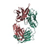
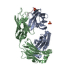
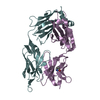
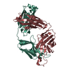
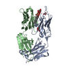
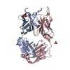
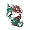
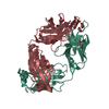
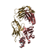
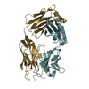
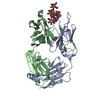
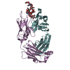
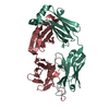
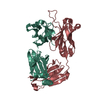
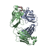
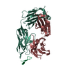
Entry Database : PDB / ID : 5eocTitle Crystal structure of Fab C2 in complex with a Cyclic variant of Hepatitis C Virus E2 epitope I (Fab fragment (Heavy chain)) x 2 ALA-CYS-GLN-LEU-ILE-ASN-THR-ASN-GLY-SER-TRP-HIS-ILE-CYS Fab fragment (Light chain) Keywords / / / Function / homology Function Domain/homology Component
/ / / / / / / / / / / / / / / / / / / / / / / / / / / / / / / / / / / / / / / / / / / / / / / / / / / / / / / / / / / / / / / / / / / / / / / / / / / / / / / / / / / / / / / / / / / / / / / / / / / / / / / / / / / / / / / / / / / / / / / / / / / / Biological species Mus musculus (house mouse)Method / / Resolution : 1.98 Å Authors Berisio, R. / Ruggiero, A. / Sandomenico, A. / Patel, A.H. / Ruvo, M. / Vitagliano, L. Funding support Organization Grant number Country Ministry of Education, Universities and Research RBNE08NKH7_003
Journal : J.Virol. / Year : 2016Title : Generation and Characterization of Monoclonal Antibodies against a Cyclic Variant of Hepatitis C Virus E2 Epitope 412-422.Authors : Sandomenico, A. / Leonardi, A. / Berisio, R. / Sanguigno, L. / Foca, G. / Foca, A. / Ruggiero, A. / Doti, N. / Muscariello, L. / Barone, D. / Farina, C. / Owsianka, A. / Vitagliano, L. / Patel, A.H. / Ruvo, M. History Deposition Nov 10, 2015 Deposition site / Processing site Revision 1.0 Feb 10, 2016 Provider / Type Revision 1.1 Mar 23, 2016 Group Revision 1.2 Jan 10, 2024 Group / Database references / Refinement descriptionCategory chem_comp_atom / chem_comp_bond ... chem_comp_atom / chem_comp_bond / database_2 / pdbx_initial_refinement_model Item / _database_2.pdbx_database_accessionRevision 1.3 Nov 20, 2024 Group / Category / pdbx_modification_feature
Show all Show less
 Movie
Movie Controller

 Yorodumi
 Open data
 Basic information
 Components
 Keywords
 Function and homology information
 Hepatitis C virus
 X-RAY DIFFRACTION /
  Authors
 Italy, 1items
  Citation
 Structure visualization
 Downloads & links
 Other downloads
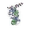


 PDBj
PDBj


 Assembly

 Mass: 18.015 Da / Num. of mol.: 476 / Source method: isolated from a natural source / Formula: H2O
 Sample preparation
 Processing