[English] 日本語
 Yorodumi
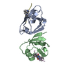
- PDB-5elu: Isoform-specific inhibition of SUMO-dependent protein-protein int... -
+ Open data
- Basic information
| Entry | Database: PDB / ID: 5elu | ||||||||||||
|---|---|---|---|---|---|---|---|---|---|---|---|---|---|
| Title | Isoform-specific inhibition of SUMO-dependent protein-protein interactions | ||||||||||||
 Components Components |
| ||||||||||||
 Keywords Keywords | SIGNALING PROTEIN / Ubiquitin / Sumoylation | ||||||||||||
| Function / homology |  Function and homology information Function and homology informationSUMO is proteolytically processed / SUMO is conjugated to E1 (UBA2:SAE1) / SUMO is transferred from E1 to E2 (UBE2I, UBC9) / Vitamin D (calciferol) metabolism / SUMOylation of SUMOylation proteins / SUMOylation of RNA binding proteins / SUMO transferase activity / SUMOylation of transcription factors / ubiquitin-like protein ligase binding / SUMOylation of DNA replication proteins ...SUMO is proteolytically processed / SUMO is conjugated to E1 (UBA2:SAE1) / SUMO is transferred from E1 to E2 (UBE2I, UBC9) / Vitamin D (calciferol) metabolism / SUMOylation of SUMOylation proteins / SUMOylation of RNA binding proteins / SUMO transferase activity / SUMOylation of transcription factors / ubiquitin-like protein ligase binding / SUMOylation of DNA replication proteins / protein sumoylation / postsynaptic cytosol / SUMOylation of DNA damage response and repair proteins / presynaptic cytosol / SUMOylation of transcription cofactors / hippocampal mossy fiber to CA3 synapse / SUMOylation of chromatin organization proteins / Regulation of endogenous retroelements by KRAB-ZFP proteins / SUMOylation of intracellular receptors / PML body / GABA-ergic synapse / protein tag activity / Formation of Incision Complex in GG-NER / positive regulation of proteasomal ubiquitin-dependent protein catabolic process / Processing of DNA double-strand break ends / ubiquitin protein ligase binding / glutamatergic synapse / positive regulation of transcription by RNA polymerase II / RNA binding / nucleoplasm / nucleus Similarity search - Function | ||||||||||||
| Biological species | synthetic construct (others) Homo sapiens (human) Homo sapiens (human) | ||||||||||||
| Method |  X-RAY DIFFRACTION / X-RAY DIFFRACTION /  SYNCHROTRON / SYNCHROTRON /  MOLECULAR REPLACEMENT / Resolution: 2.35 Å MOLECULAR REPLACEMENT / Resolution: 2.35 Å | ||||||||||||
 Authors Authors | Hughes, D.J. / Tiede, C. / Hall, N. / Tang, A.A.S. / Trinh, C.H. / Zajac, K. / Mandal, U. / Howell, G. / Edwards, T.A. / McPherson, M.J. ...Hughes, D.J. / Tiede, C. / Hall, N. / Tang, A.A.S. / Trinh, C.H. / Zajac, K. / Mandal, U. / Howell, G. / Edwards, T.A. / McPherson, M.J. / Tomlinson, D.C. / Whitehouse, A. | ||||||||||||
| Funding support |  United Kingdom, 3items United Kingdom, 3items
| ||||||||||||
 Citation Citation |  Journal: Sci Signal / Year: 2017 Journal: Sci Signal / Year: 2017Title: Generation of specific inhibitors of SUMO-1- and SUMO-2/3-mediated protein-protein interactions using Affimer (Adhiron) technology. Authors: Hughes, D.J. / Tiede, C. / Penswick, N. / Tang, A.A. / Trinh, C.H. / Mandal, U. / Zajac, K.Z. / Gaule, T. / Howell, G. / Edwards, T.A. / Duan, J. / Feyfant, E. / McPherson, M.J. / Tomlinson, ...Authors: Hughes, D.J. / Tiede, C. / Penswick, N. / Tang, A.A. / Trinh, C.H. / Mandal, U. / Zajac, K.Z. / Gaule, T. / Howell, G. / Edwards, T.A. / Duan, J. / Feyfant, E. / McPherson, M.J. / Tomlinson, D.C. / Whitehouse, A. | ||||||||||||
| History |
|
- Structure visualization
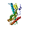
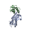
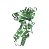
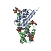
| Structure viewer | Molecule:  Molmil Molmil Jmol/JSmol Jmol/JSmol |
|---|
- Downloads & links
- Download
| PDBx/mmCIF format |  5elu.cif.gz 5elu.cif.gz | 51.5 KB | Display |  PDBx/mmCIF format PDBx/mmCIF format |
|---|---|---|---|---|
| PDB format |  pdb5elu.ent.gz pdb5elu.ent.gz | 34.3 KB | Display |  PDB format PDB format |
| PDBx/mmJSON format |  5elu.json.gz 5elu.json.gz | Tree view |  PDBx/mmJSON format PDBx/mmJSON format | |
| Others |  Other downloads Other downloads |
-Validation report
| Arichive directory |  https://data.pdbj.org/pub/pdb/validation_reports/el/5elu https://data.pdbj.org/pub/pdb/validation_reports/el/5elu ftp://data.pdbj.org/pub/pdb/validation_reports/el/5elu ftp://data.pdbj.org/pub/pdb/validation_reports/el/5elu | HTTPS FTP |
|---|
-Related structure data
| Related structure data |  5eljSC  5eqlC  1wm3S S: Starting model for refinement C: citing same article ( |
|---|---|
| Similar structure data |
- Links
Links
- Assembly
| Deposited unit | 
| ||||||||
|---|---|---|---|---|---|---|---|---|---|
| 1 |
| ||||||||
| Unit cell |
|
- Components
| #1: Protein | Mass: 13509.390 Da / Num. of mol.: 1 Source method: isolated from a genetically manipulated source Details: MASAATGVRAVPGNENSLEIEELARFAVDEHNKKENALLEFVRVVKAKEQVDLTRFPVTTMYYLTLEAKDGGKKKLYEAKVWVKGYLLEELKHNFKELQEFKPVGDAAAAHHHHHHHH Source: (gene. exp.) synthetic construct (others) / Gene: PHYTOCYSTATIN / Plasmid: PET11 / Production host:  | ||
|---|---|---|---|
| #2: Protein | Mass: 8965.129 Da / Num. of mol.: 1 Source method: isolated from a genetically manipulated source Source: (gene. exp.)  Homo sapiens (human) / Gene: SUMO2, SMT3B, SMT3H2 / Plasmid: PET11 / Production host: Homo sapiens (human) / Gene: SUMO2, SMT3B, SMT3H2 / Plasmid: PET11 / Production host:  | ||
| #3: Chemical | | #4: Water | ChemComp-HOH / | |
-Experimental details
-Experiment
| Experiment | Method:  X-RAY DIFFRACTION X-RAY DIFFRACTION |
|---|
- Sample preparation
| Crystal | Density Matthews: 2.24 Å3/Da / Density % sol: 45.12 % |
|---|---|
| Crystal grow | Temperature: 291 K / Method: vapor diffusion, sitting drop / pH: 6.5 Details: 0.1 M sodium cacodylate pH 6.5, 0.2 M sodium chloride and 2.0 M ammonium sulphate |
-Data collection
| Diffraction | Mean temperature: 100 K |
|---|---|
| Diffraction source | Source:  SYNCHROTRON / Site: SYNCHROTRON / Site:  Diamond Diamond  / Beamline: I03 / Wavelength: 0.98 Å / Beamline: I03 / Wavelength: 0.98 Å |
| Detector | Type: DECTRIS PILATUS 6M / Detector: PIXEL / Date: Sep 26, 2014 |
| Radiation | Monochromator: SAGITALLY FOCUSED Si (111) / Protocol: SINGLE WAVELENGTH / Monochromatic (M) / Laue (L): M / Scattering type: x-ray |
| Radiation wavelength | Wavelength: 0.98 Å / Relative weight: 1 |
| Reflection | Resolution: 2.35→28.4 Å / Num. obs: 8893 / % possible obs: 99.6 % / Observed criterion σ(F): 2 / Observed criterion σ(I): 2 / Redundancy: 5.9 % / Biso Wilson estimate: 19 Å2 / Rmerge(I) obs: 0.043 / Net I/σ(I): 29.4 |
| Reflection shell | Resolution: 2.35→2.44 Å / Redundancy: 4.3 % / Rmerge(I) obs: 0.11 / Mean I/σ(I) obs: 11.1 / % possible all: 96.9 |
- Processing
| Software |
| ||||||||||||||||||||||||||||||||||||||||||
|---|---|---|---|---|---|---|---|---|---|---|---|---|---|---|---|---|---|---|---|---|---|---|---|---|---|---|---|---|---|---|---|---|---|---|---|---|---|---|---|---|---|---|---|
| Refinement | Method to determine structure:  MOLECULAR REPLACEMENT MOLECULAR REPLACEMENTStarting model: PDB ENTRIES 1WM3 and 5ELJ Resolution: 2.35→28.393 Å / SU ML: 0.26 / Cross valid method: FREE R-VALUE / σ(F): 0.79 / Phase error: 0.217 / Stereochemistry target values: ML
| ||||||||||||||||||||||||||||||||||||||||||
| Solvent computation | Shrinkage radii: 0.9 Å / VDW probe radii: 1.11 Å / Solvent model: FLAT BULK SOLVENT MODEL | ||||||||||||||||||||||||||||||||||||||||||
| Refinement step | Cycle: LAST / Resolution: 2.35→28.393 Å
| ||||||||||||||||||||||||||||||||||||||||||
| Refine LS restraints |
| ||||||||||||||||||||||||||||||||||||||||||
| LS refinement shell |
|
 Movie
Movie Controller




 PDBj
PDBj


