- PDB-1wm3: Crystal structure of human SUMO-2 protein -
+
Open data
ID or keywords:
Loading...
-
Basic information
Entry
Database: PDB / ID: 1wm3
Title
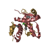
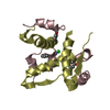
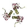
Crystal structure of human SUMO-2 protein
Components
Ubiquitin-like protein SMT3B
Keywords
PROTEIN TRANSPORT / ubiquitin fold / half-open barrel / two helices
Function / homology
Function and homology information
SUMO is proteolytically processed / SUMO is conjugated to E1 (UBA2:SAE1) / SUMO is transferred from E1 to E2 (UBE2I, UBC9) / Vitamin D (calciferol) metabolism / SUMOylation of SUMOylation proteins / SUMOylation of RNA binding proteins / SUMO transferase activity / SUMOylation of transcription factors / SUMOylation of DNA replication proteins / ubiquitin-like protein ligase binding ...SUMO is proteolytically processed / SUMO is conjugated to E1 (UBA2:SAE1) / SUMO is transferred from E1 to E2 (UBE2I, UBC9) / Vitamin D (calciferol) metabolism / SUMOylation of SUMOylation proteins / SUMOylation of RNA binding proteins / SUMO transferase activity / SUMOylation of transcription factors / SUMOylation of DNA replication proteins / ubiquitin-like protein ligase binding / protein sumoylation / postsynaptic cytosol / SUMOylation of DNA damage response and repair proteins / presynaptic cytosol / SUMOylation of transcription cofactors / SUMOylation of chromatin organization proteins / hippocampal mossy fiber to CA3 synapse / Regulation of endogenous retroelements by KRAB-ZFP proteins / SUMOylation of intracellular receptors / PML body / GABA-ergic synapse / Formation of Incision Complex in GG-NER / protein tag activity / positive regulation of proteasomal ubiquitin-dependent protein catabolic process / Processing of DNA double-strand break ends / nuclear body / ubiquitin protein ligase binding / glutamatergic synapse / RNA binding / nucleoplasm / nucleus Similarity search - Function
In the structure databanks used in Yorodumi, some data are registered as the other names, "COVID-19 virus" and "2019-nCoV". Here are the details of the virus and the list of structure data.
Jan 31, 2019. EMDB accession codes are about to change! (news from PDBe EMDB page)
EMDB accession codes are about to change! (news from PDBe EMDB page)
The allocation of 4 digits for EMDB accession codes will soon come to an end. Whilst these codes will remain in use, new EMDB accession codes will include an additional digit and will expand incrementally as the available range of codes is exhausted. The current 4-digit format prefixed with “EMD-” (i.e. EMD-XXXX) will advance to a 5-digit format (i.e. EMD-XXXXX), and so on. It is currently estimated that the 4-digit codes will be depleted around Spring 2019, at which point the 5-digit format will come into force.
The EM Navigator/Yorodumi systems omit the EMD- prefix.
Related info.:Q: What is EMD? / ID/Accession-code notation in Yorodumi/EM Navigator
Yorodumi is a browser for structure data from EMDB, PDB, SASBDB, etc.
This page is also the successor to EM Navigator detail page, and also detail information page/front-end page for Omokage search.
The word "yorodu" (or yorozu) is an old Japanese word meaning "ten thousand". "mi" (miru) is to see.
Related info.:EMDB / PDB / SASBDB / Comparison of 3 databanks / Yorodumi Search / Aug 31, 2016. New EM Navigator & Yorodumi / Yorodumi Papers / Jmol/JSmol / Function and homology information / Changes in new EM Navigator and Yorodumi
 Movie
Movie Controller


 Open data
 Basic information
 Components
 Keywords
 Function and homology information
 Homo sapiens (human)
 X-RAY DIFFRACTION /
  Authors
 Citation
 Structure visualization
 Downloads & links
 Other downloads


 PDBj
PDBj


 Assembly


 Mass: 18.015 Da / Num. of mol.: 127 / Source method: isolated from a natural source / Formula: H2O
 Sample preparation
 / Beamline: BL17B2 / Wavelength: 1.0717 Å
 Processing