 Movie
Movie Controller


+ Open data

- Basic information
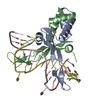
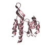
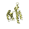
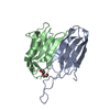
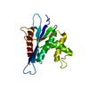
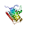
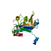
| Entry | Database: PDB / ID: 5eka | ||||||
|---|---|---|---|---|---|---|---|
| Title | HU DNA-binding protein from Thermus thermophilus | ||||||
 Components Components | DNA-binding protein HU | ||||||
 Keywords Keywords | DNA BINDING PROTEIN / HU PROTEIN / HISTONE-LIKE PROTEIN / THERMOSTABLE DNA-BINDING PROTEIN | ||||||
| Function / homology |  Function and homology information Function and homology informationchromosome condensation / structural constituent of chromatin / DNA binding / cytosol Similarity search - Function | ||||||
| Biological species |   Thermus thermophilus HB8 (bacteria) Thermus thermophilus HB8 (bacteria) | ||||||
| Method |  X-RAY DIFFRACTION / SYNCHROTRON / MOLECULAR REPLACEMENT / Resolution: 1.69 Å X-RAY DIFFRACTION / SYNCHROTRON / MOLECULAR REPLACEMENT / Resolution: 1.69 Å | ||||||
 Authors Authors | Papageorgiou, A. / Adam, P. / Stavros, P. / Nounesis, G. / Meijers, R. / Petratos, K. / Vorgias, C.E. | ||||||
 Citation Citation | Journal: Extremophiles / Year: 2016 Title: HU histone-like DNA-binding protein from Thermus thermophilus: structural and evolutionary analyses. Authors: Papageorgiou, A.C. / Adam, P.S. / Stavros, P. / Nounesis, G. / Meijers, R. / Petratos, K. / Vorgias, C.E. | ||||||
| History |
|
- Structure visualization
| Structure viewer | Molecule: MolmilJmol/JSmol |
|---|
- Downloads & links
-Download
| PDBx/mmCIF format | 5eka.cif.gz | 49.4 KB | Display | PDBx/mmCIF format |
|---|---|---|---|---|
| PDB format | pdb5eka.ent.gz | 34.9 KB | Display | PDB format |
| PDBx/mmJSON format | 5eka.json.gz | Tree view | PDBx/mmJSON format | |
| Others |  Other downloads Other downloads |
-Validation report
| Arichive directory | https://data.pdbj.org/pub/pdb/validation_reports/ek/5ekaftp://data.pdbj.org/pub/pdb/validation_reports/ek/5eka | HTTPS FTP |
|---|
-Related structure data
| Related structure data |  1b8zS S: Starting model for refinement |
|---|---|
| Similar structure data |


-Links
 PDBj
PDBj- Assembly
| Deposited unit | 
| ||||||||
|---|---|---|---|---|---|---|---|---|---|
| 1 | 
| ||||||||
| Unit cell |
| ||||||||
| Components on special symmetry positions |
|
-Components
| #1: Protein | Mass: 10345.323 Da / Num. of mol.: 1 Source method: isolated from a genetically manipulated source Details: DNA-binding protein HU from Thermus thermophilus. UniProtKB code: DBH_THET8 Source: (gene. exp.) Thermus thermophilus HB8 (bacteria) / Gene: TTHA1349 / Production host: |
|---|---|
| #2: Chemical | ChemComp-GOL /   Mass: 92.094 Da / Num. of mol.: 1 / Source method: obtained synthetically / Formula: C3H8O3 Mass: 92.094 Da / Num. of mol.: 1 / Source method: obtained synthetically / Formula: C3H8O3 |
| #3: Water | ChemComp-HOH /  Mass: 18.015 Da / Num. of mol.: 96 / Source method: isolated from a natural source / Formula: H2O Mass: 18.015 Da / Num. of mol.: 96 / Source method: isolated from a natural source / Formula: H2O |
-Experimental details
-Experiment
| Experiment | Method: X-RAY DIFFRACTION |
|---|
- Sample preparation
| Crystal | Density Matthews: 2.3 Å3/Da / Density % sol: 51 % / Description: Rod-shaped crystals |
|---|---|
| Crystal grow | Temperature: 293 K / Method: vapor diffusion, hanging drop / pH: 4.3 Details: Protein solutions of 14-18 mg/ml, Buffer: 0.2 M Na-formate, Precipitating agent: 20%(w/v) PEG 3350 |
-Data collection
| Diffraction | Mean temperature: 100 K |
|---|---|
| Diffraction source | Source: SYNCHROTRON / Site: PETRA III, EMBL c/o DESY  / Beamline: P14 (MX2) / Wavelength: 0.97631 Å / Beamline: P14 (MX2) / Wavelength: 0.97631 Å |
| Detector | Type: DECTRIS PILATUS 6M / Detector: PIXEL / Date: Apr 26, 2015 / Details: BIMORPH MIRRORS |
| Radiation | Monochromator: Si111 / Protocol: SINGLE WAVELENGTH / Monochromatic (M) / Laue (L): M / Scattering type: x-ray |
| Radiation wavelength | Wavelength: 0.97631 Å / Relative weight: 1 |
| Reflection | Resolution: 1.69→44.9 Å / Num. obs: 10742 / % possible obs: 96.3 % / Redundancy: 12 % / Biso Wilson estimate: 20.4 Å2 / Rmerge(I) obs: 0.073 / Net I/av σ(I): 17.6 / Net I/σ(I): 22.6 |
| Reflection shell | Resolution: 1.69→1.74 Å / Redundancy: 7.2 % / Rmerge(I) obs: 0.51 / Mean I/σ(I) obs: 3 / % possible all: 59.6 |
- Processing
| Software |
| ||||||||||||||||||||||||||||||||||||||||||||||||||||||||||||||||||||||||||||||||||||||||||||||||||||||||||||||||||||||||||||||||||||||||||||||||||||||||||||||||||||||||||||||||||||||
|---|---|---|---|---|---|---|---|---|---|---|---|---|---|---|---|---|---|---|---|---|---|---|---|---|---|---|---|---|---|---|---|---|---|---|---|---|---|---|---|---|---|---|---|---|---|---|---|---|---|---|---|---|---|---|---|---|---|---|---|---|---|---|---|---|---|---|---|---|---|---|---|---|---|---|---|---|---|---|---|---|---|---|---|---|---|---|---|---|---|---|---|---|---|---|---|---|---|---|---|---|---|---|---|---|---|---|---|---|---|---|---|---|---|---|---|---|---|---|---|---|---|---|---|---|---|---|---|---|---|---|---|---|---|---|---|---|---|---|---|---|---|---|---|---|---|---|---|---|---|---|---|---|---|---|---|---|---|---|---|---|---|---|---|---|---|---|---|---|---|---|---|---|---|---|---|---|---|---|---|---|---|---|---|
| Refinement | Method to determine structure: MOLECULAR REPLACEMENT Starting model: 1B8Z Resolution: 1.69→44.86 Å / Cor.coef. Fo:Fc: 0.946 / Cor.coef. Fo:Fc free: 0.909 / SU B: 4.387 / SU ML: 0.077 / Cross valid method: THROUGHOUT / ESU R: 0.112 / ESU R Free: 0.122 / Stereochemistry target values: MAXIMUM LIKELIHOOD / Details: HYDROGENS HAVE BEEN ADDED IN THE RIDING POSITIONS
| ||||||||||||||||||||||||||||||||||||||||||||||||||||||||||||||||||||||||||||||||||||||||||||||||||||||||||||||||||||||||||||||||||||||||||||||||||||||||||||||||||||||||||||||||||||||
| Solvent computation | Ion probe radii: 0.8 Å / Shrinkage radii: 0.8 Å / VDW probe radii: 1.2 Å / Solvent model: BABINET MODEL WITH MASK | ||||||||||||||||||||||||||||||||||||||||||||||||||||||||||||||||||||||||||||||||||||||||||||||||||||||||||||||||||||||||||||||||||||||||||||||||||||||||||||||||||||||||||||||||||||||
| Displacement parameters | Biso mean: 31 Å2
| ||||||||||||||||||||||||||||||||||||||||||||||||||||||||||||||||||||||||||||||||||||||||||||||||||||||||||||||||||||||||||||||||||||||||||||||||||||||||||||||||||||||||||||||||||||||
| Refinement step | Cycle: 1 / Resolution: 1.69→44.86 Å
| ||||||||||||||||||||||||||||||||||||||||||||||||||||||||||||||||||||||||||||||||||||||||||||||||||||||||||||||||||||||||||||||||||||||||||||||||||||||||||||||||||||||||||||||||||||||
| Refine LS restraints |
|