 Movie
Movie Controller

[English] 日本語
 Yorodumi
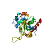
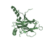
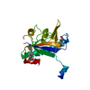
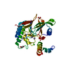
- PDB-6x8z: Crystal structure of N-truncated human B12 chaperone CblD(C262S)-... -
+ Open data

- Basic information
| Entry | Database: PDB / ID: 6x8z | ||||||
|---|---|---|---|---|---|---|---|
| Title | Crystal structure of N-truncated human B12 chaperone CblD(C262S)-thiolato-cob(III)alamin complex (108-296) | ||||||
 Components Components | Methylmalonic aciduria and homocystinuria type D protein, mitochondrial | ||||||
 Keywords Keywords | OXIDOREDUCTASE / cobalamin / vitamin B12 / chaperone | ||||||
| Function / homology |  Function and homology information Function and homology informationDefective MMADHC causes MMAHCD / Cobalamin (Cbl) metabolism / cobalamin metabolic process / molecular carrier activity / mitochondrion / cytoplasm / cytosol Similarity search - Function | ||||||
| Biological species |  Homo sapiens (human) Homo sapiens (human) | ||||||
| Method |  X-RAY DIFFRACTION / SYNCHROTRON / MOLECULAR REPLACEMENT / Resolution: 2.5 Å X-RAY DIFFRACTION / SYNCHROTRON / MOLECULAR REPLACEMENT / Resolution: 2.5 Å | ||||||
 Authors Authors | Mascarenhas, R. / Li, Z. / Koutmos, M. / Banerjee, R. | ||||||
| Funding support |  United States, 1items United States, 1items
| ||||||
 Citation Citation | Journal: J.Am.Chem.Soc. / Year: 2020 Title: An Interprotein Co-S Coordination Complex in the B 12 -Trafficking Pathway. Authors: Li, Z. / Mascarenhas, R. / Twahir, U.T. / Kallon, A. / Deb, A. / Yaw, M. / Penner-Hahn, J. / Koutmos, M. / Warncke, K. / Banerjee, R. | ||||||
| History |
|
- Structure visualization
| Structure viewer | Molecule: MolmilJmol/JSmol |
|---|
- Downloads & links
-Download
| PDBx/mmCIF format | 6x8z.cif.gz | 133.3 KB | Display | PDBx/mmCIF format |
|---|---|---|---|---|
| PDB format | pdb6x8z.ent.gz | 104.4 KB | Display | PDB format |
| PDBx/mmJSON format | 6x8z.json.gz | Tree view | PDBx/mmJSON format | |
| Others |  Other downloads Other downloads |
-Validation report
| Arichive directory | https://data.pdbj.org/pub/pdb/validation_reports/x8/6x8zftp://data.pdbj.org/pub/pdb/validation_reports/x8/6x8z | HTTPS FTP |
|---|
-Related structure data
| Related structure data |  5cv0S S: Starting model for refinement |
|---|---|
| Similar structure data |


-Links
 PDBj
PDBj
- Assembly
| Deposited unit | 
| ||||||||
|---|---|---|---|---|---|---|---|---|---|
| 1 | 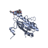
| ||||||||
| 2 | 
| ||||||||
| Unit cell |
|
-Components
| #1: Protein | Mass: 21824.441 Da / Num. of mol.: 2 / Fragment: UNP residues 108-296 / Mutation: C262S Source method: isolated from a genetically manipulated source Source: (gene. exp.) Homo sapiens (human) / Gene: MMADHC, C2orf25, CL25022, HSPC161, My011 / Production host:  #2: Chemical |   Mass: 1330.356 Da / Num. of mol.: 2 / Source method: obtained synthetically / Formula: C62H89CoN13O14P / Feature type: SUBJECT OF INVESTIGATION Mass: 1330.356 Da / Num. of mol.: 2 / Source method: obtained synthetically / Formula: C62H89CoN13O14P / Feature type: SUBJECT OF INVESTIGATION#3: Water | ChemComp-HOH / |  Mass: 18.015 Da / Num. of mol.: 14 / Source method: isolated from a natural source / Formula: H2O Mass: 18.015 Da / Num. of mol.: 14 / Source method: isolated from a natural source / Formula: H2OHas ligand of interest | Y | |
|---|
-Experimental details
-Experiment
| Experiment | Method: X-RAY DIFFRACTION / Number of used crystals: 1 |
|---|
- Sample preparation
| Crystal | Density Matthews: 2.6 Å3/Da / Density % sol: 52.71 % |
|---|---|
| Crystal grow | Temperature: 293 K / Method: vapor diffusion, hanging drop Details: 20% PEG8000, 100 mM Tris-HCl, pH 8.5, 200 mM magnesium chloride |
-Data collection
| Diffraction | Mean temperature: 100 K / Serial crystal experiment: N |
|---|---|
| Diffraction source | Source: SYNCHROTRON / Site: APS / Beamline: 21-ID-G / Wavelength: 0.987 Å |
| Detector | Type: MARMOSAIC 300 mm CCD / Detector: CCD / Date: Jun 6, 2018 |
| Radiation | Monochromator: diamond(111) / Protocol: SINGLE WAVELENGTH / Monochromatic (M) / Laue (L): M / Scattering type: x-ray |
| Radiation wavelength | Wavelength: 0.987 Å / Relative weight: 1 |
| Reflection | Resolution: 2.5→50.637 Å / Num. obs: 15518 / % possible obs: 99.4 % / Redundancy: 4.1 % / CC1/2: 0.993 / Rmerge(I) obs: 0.107 / Net I/σ(I): 7.6 |
| Reflection shell | Resolution: 2.5→2.6 Å / Rmerge(I) obs: 1.213 / Num. unique obs: 1736 / CC1/2: 0.593 |
- Processing
| Software |
| |||||||||||||||||||||||||||||||||||
|---|---|---|---|---|---|---|---|---|---|---|---|---|---|---|---|---|---|---|---|---|---|---|---|---|---|---|---|---|---|---|---|---|---|---|---|---|
| Refinement | Method to determine structure: MOLECULAR REPLACEMENT Starting model: PDB entry 5CV0 Resolution: 2.5→50.637 Å / SU ML: 0.38 / Cross valid method: THROUGHOUT / σ(F): 1.35 / Phase error: 33.64 / Stereochemistry target values: ML
| |||||||||||||||||||||||||||||||||||
| Solvent computation | Shrinkage radii: 0.9 Å / VDW probe radii: 1.11 Å / Solvent model: FLAT BULK SOLVENT MODEL | |||||||||||||||||||||||||||||||||||
| Displacement parameters | Biso max: 136.93 Å2 / Biso mean: 64.5675 Å2 / Biso min: 30.6 Å2 | |||||||||||||||||||||||||||||||||||
| Refinement step | Cycle: final / Resolution: 2.5→50.637 Å
| |||||||||||||||||||||||||||||||||||
| LS refinement shell | Refine-ID: X-RAY DIFFRACTION / Rfactor Rfree error: 0 / % reflection obs: 99 %
|
