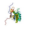
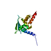
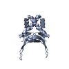
THE ASYMMETRIC UNIT CONSISTS OF THREE HU MONOMERS ARRANGED AROUND THREE OF THE FOUR TWOFOLD AXES IN THE P2 SPACE GROUP. BIOLOGICALLY RELEVANT DIMERS ARE GENERATED BY THE SYMMETRY OPERATIONS OF THE UNIT CELL. THE SOLVENT MOLECULES ARE LABELED ACCORDING TO THE MONOMER WITH WHICH THEY ASSOCIATE. WATER MOLECULES 100 THROUGH 135 ARE COMMON TO ALL THREE MOLECULES IN THE ASYMMETRIC UNIT AND CAN BE CONSIDERED STRUCTURAL. WATER MOLECULES 200 AND HIGHER ARE NOT COMMON TO ALL THREE MOLECULES.
-
Components
#1: Protein
PROTEINHU / BSB / NS
Mass: 9733.168 Da / Num. of mol.: 3 / Source method: isolated from a natural source / Source: (natural) Geobacillus stearothermophilus (bacteria) / References: UniProt: P02346, UniProt: P0A3H0*PLUS
Mass: 18.015 Da / Num. of mol.: 271 / Source method: isolated from a natural source / Formula: H2O
Compound details
PROTEIN HU BINDS DNA NON-SPECIFICALLY AND INTRODUCES SHARP BENDS. THE PROTEIN APPEARS TO INDUCE DNA ...PROTEIN HU BINDS DNA NON-SPECIFICALLY AND INTRODUCES SHARP BENDS. THE PROTEIN APPEARS TO INDUCE DNA NEGATIVE SUPERCOILING BY PROTEIN-PROTEIN INTERACTION. BIOLOGICAL ROLE IS TO INDUCE DNA SUPERCOILING AND RELIEVE TORSIONAL STRESS RESULTING FROM DNA PROCESSES SUCH AS TRANSCRIPTION AND REPLICATION.
-
Experimental details
-
Experiment
Experiment
Method: X-RAY DIFFRACTION / Number of used crystals: 2
-
Sample preparation
Crystal
Density Matthews: 2.12 Å3/Da / Density % sol: 51 %
Crystal grow
pH: 8 / Details: pH 8.0
Crystal grow
*PLUS
Method: unknown / PH range low: 9 / PH range high: 7
Resolution: 2→10 Å / Num. obs: 18328 / % possible obs: 89 % / Observed criterion σ(I): 2 / Redundancy: 4 % / Biso Wilson estimate: 27 Å2 / Rmerge(I) obs: 0.07 / Rsym value: 0.07 / Net I/σ(I): 10
Reflection shell
Resolution: 2→2.19 Å / Redundancy: 2 % / Rmerge(I) obs: 0.1 / Mean I/σ(I) obs: 2.5 / Rsym value: 0.1 / % possible all: 80
Reflection shell
*PLUS
% possible obs: 80 %
-
Processing
Software
Name
Version
Classification
X-PLOR
3.8
modelbuilding
X-PLOR
3.8
refinement
MOSFLM
datareduction
CCP4
datascaling
X-PLOR
3.8
phasing
Refinement
Method to determine structure: SINGLE ISOMORPHOUS REPLACEMENT PLUS ANOMALOUS (URANYL), NCS AVERAGING ON THREE MOLECULES Resolution: 2→6 Å / Rfactor Rfree error: 0.017 / Data cutoff high absF: 1000000 / Data cutoff low absF: 0.001 / Cross valid method: THROUGHOUT / σ(F): 2 Details: THE MOLECULE CONTAINS A DISORDERED ARM REGION THAT IS KNOWN TO BIND DNA IN THE MINOR GROOVE FROM THE RELATED IHF STRUCTURE. EACH OF THE THREE MOLECULES IN THE ASYMMETRIC UNIT IS MISSING ...Details: THE MOLECULE CONTAINS A DISORDERED ARM REGION THAT IS KNOWN TO BIND DNA IN THE MINOR GROOVE FROM THE RELATED IHF STRUCTURE. EACH OF THE THREE MOLECULES IN THE ASYMMETRIC UNIT IS MISSING DIFFERENT AMOUNTS OF THE ARM DUE TO THE LACK OF ELECTRON DENSITY. MOLECULE A IS MISSING 59 THROUGH 68, MOLECULE B 57 THROUGH 72, AND MOLECULE C 56 THROUGH 74.
Rfactor
Num. reflection
% reflection
Selection details
Rfree
0.213
333
10 %
RANDOM
Rwork
0.193
-
-
-
obs
0.193
17251
89 %
-
Displacement parameters
Biso mean: 27 Å2
Baniso -1
Baniso -2
Baniso -3
1-
0 Å2
0 Å2
0 Å2
2-
-
0 Å2
0 Å2
3-
-
-
0 Å2
Refinement step
Cycle: LAST / Resolution: 2→6 Å
Protein
Nucleic acid
Ligand
Solvent
Total
Num. atoms
1700
0
0
271
1971
Refine LS restraints
Refine-ID
Type
Dev ideal
X-RAY DIFFRACTION
x_bond_d
0.011
X-RAY DIFFRACTION
x_bond_d_na
X-RAY DIFFRACTION
x_bond_d_prot
X-RAY DIFFRACTION
x_angle_d
X-RAY DIFFRACTION
x_angle_d_na
X-RAY DIFFRACTION
x_angle_d_prot
X-RAY DIFFRACTION
x_angle_deg
1.53
X-RAY DIFFRACTION
x_angle_deg_na
X-RAY DIFFRACTION
x_angle_deg_prot
X-RAY DIFFRACTION
x_dihedral_angle_d
24.6
X-RAY DIFFRACTION
x_dihedral_angle_d_na
X-RAY DIFFRACTION
x_dihedral_angle_d_prot
X-RAY DIFFRACTION
x_improper_angle_d
1.6
X-RAY DIFFRACTION
x_improper_angle_d_na
X-RAY DIFFRACTION
x_improper_angle_d_prot
X-RAY DIFFRACTION
x_mcbond_it
X-RAY DIFFRACTION
x_mcangle_it
X-RAY DIFFRACTION
x_scbond_it
X-RAY DIFFRACTION
x_scangle_it
LS refinement shell
Resolution: 2→2.19 Å / Rfactor Rfree error: 0.04 / Total num. of bins used: 8
Rfactor
Num. reflection
% reflection
Rfree
0.33
55
12 %
Rwork
0.289
1743
-
obs
-
-
80 %
Xplor file
Refine-ID
Serial no
Param file
Topol file
X-RAY DIFFRACTION
1
PARAM19X.PRO
TOPH19X.PRO
X-RAY DIFFRACTION
2
+
About Yorodumi
-
News
-
Feb 9, 2022. New format data for meta-information of EMDB entries
New format data for meta-information of EMDB entries
Version 3 of the EMDB header file is now the official format.
The previous official version 1.9 will be removed from the archive.
In the structure databanks used in Yorodumi, some data are registered as the other names, "COVID-19 virus" and "2019-nCoV". Here are the details of the virus and the list of structure data.
Jan 31, 2019. EMDB accession codes are about to change! (news from PDBe EMDB page)
EMDB accession codes are about to change! (news from PDBe EMDB page)
The allocation of 4 digits for EMDB accession codes will soon come to an end. Whilst these codes will remain in use, new EMDB accession codes will include an additional digit and will expand incrementally as the available range of codes is exhausted. The current 4-digit format prefixed with “EMD-” (i.e. EMD-XXXX) will advance to a 5-digit format (i.e. EMD-XXXXX), and so on. It is currently estimated that the 4-digit codes will be depleted around Spring 2019, at which point the 5-digit format will come into force.
The EM Navigator/Yorodumi systems omit the EMD- prefix.
Related info.:Q: What is EMD? / ID/Accession-code notation in Yorodumi/EM Navigator
Yorodumi is a browser for structure data from EMDB, PDB, SASBDB, etc.
This page is also the successor to EM Navigator detail page, and also detail information page/front-end page for Omokage search.
The word "yorodu" (or yorozu) is an old Japanese word meaning "ten thousand". "mi" (miru) is to see.
Related info.:EMDB / PDB / SASBDB / Comparison of 3 databanks / Yorodumi Search / Aug 31, 2016. New EM Navigator & Yorodumi / Yorodumi Papers / Jmol/JSmol / Function and homology information / Changes in new EM Navigator and Yorodumi
 Movie
Movie Controller


 Open data
 Basic information
 Components
 Keywords
 Function and homology information
 Geobacillus stearothermophilus (bacteria)
 X-RAY DIFFRACTION /
  Authors
 Citation
 Structure visualization
 Downloads & links
 Other downloads
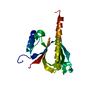


 PDBj
PDBj Assembly
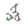


 Mass: 18.015 Da / Num. of mol.: 271 / Source method: isolated from a natural source / Formula: H2O
 Sample preparation
 / Beamline: PX7.2 / Wavelength: 1.488
  Processing