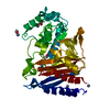
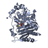
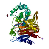
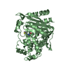
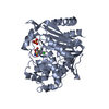
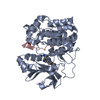
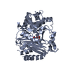
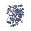
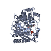
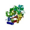
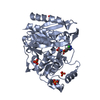
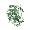
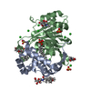
CRYSTAL STRUCTURE OF Fox-4 cephamycinase mutant Y150F complexed with cefoxitin
Components
Beta-lactamase
Keywords
HYDROLASE / beta-lactamase
Function / homology
Function and homology information
antibiotic catabolic process / beta-lactamase activity / beta-lactamase / outer membrane-bounded periplasmic space / response to antibiotic / metal ion binding Similarity search - Function
Protocol: SINGLE WAVELENGTH / Monochromatic (M) / Laue (L): M / Scattering type: x-ray
Radiation wavelength
Wavelength: 0.9791 Å / Relative weight: 1
Reflection
Redundancy: 3.7 % / Number: 355116 / Rmerge(I) obs: 0.07 / Rsym value: 0.07 / D res high: 1.21 Å / D res low: 19.676 Å / Num. obs: 96864 / % possible obs: 95.4
Diffraction reflection shell
Highest resolution (Å)
Lowest resolution (Å)
ID
Rmerge(I) obs
Rsym value
Redundancy
3.83
19.68
1
0.033
0.033
3.6
2.71
3.83
1
0.037
0.037
3.8
2.21
2.71
1
0.057
0.057
3.8
1.91
2.21
1
0.081
0.081
3.8
1.71
1.91
1
0.105
0.105
3.8
1.56
1.71
1
0.139
0.139
3.7
1.45
1.56
1
0.218
0.218
3.6
1.35
1.45
1
0.36
0.36
3.6
1.28
1.35
1
0.508
0.508
3.6
1.21
1.28
1
0.739
0.739
3.6
Reflection
Resolution: 1.21→19.676 Å / Num. obs: 96864 / % possible obs: 95.4 % / Redundancy: 3.7 % / Biso Wilson estimate: 11.41 Å2 / Rmerge(I) obs: 0.07 / Rsym value: 0.07 / Net I/av σ(I): 7.16 / Net I/σ(I): 6.4 / Num. measured all: 355116
Reflection shell
Diffraction-ID: 1 / Rejects: _
Resolution (Å)
Redundancy (%)
Rmerge(I) obs
Mean I/σ(I) obs
Num. measured all
Num. unique all
Rsym value
Net I/σ(I) obs
% possible all
1.21-1.28
3.6
0.739
1
48683
13617
0.739
1.6
92.2
1.28-1.35
3.6
0.508
1.5
46623
13059
0.508
2.3
93.2
1.35-1.45
3.6
0.36
2.1
44640
12416
0.36
3.2
94.6
1.45-1.56
3.6
0.218
3.5
42478
11659
0.218
4.9
95.5
1.56-1.71
3.7
0.139
5.3
40388
10870
0.139
6.8
96.4
1.71-1.91
3.8
0.105
6.5
37417
9945
0.105
8.6
97.4
1.91-2.21
3.8
0.081
7.9
33308
8814
0.081
11
97.8
2.21-2.71
3.8
0.057
11.1
28448
7539
0.057
12.5
98.4
2.71-3.83
3.8
0.037
16.3
21955
5843
0.037
13.6
98.6
3.83-19.676
3.6
0.033
18.4
11176
3102
0.033
13.5
93.4
-
Phasing
Phasing
Method: molecular replacement
Phasing MR
Model details: Phaser MODE: MR_AUTO
Highest resolution
Lowest resolution
Rotation
2.5 Å
19.64 Å
Translation
2.5 Å
19.64 Å
-
Processing
Software
Name
Version
Classification
PHASER
2.5.6
phasing
PHENIX
refinement
PDB_EXTRACT
3.15
dataextraction
MOSFLM
datareduction
SCALA
datascaling
PHASER
phasing
Refinement
Method to determine structure: MOLECULAR REPLACEMENT Starting model: PDB 5CGS
In the structure databanks used in Yorodumi, some data are registered as the other names, "COVID-19 virus" and "2019-nCoV". Here are the details of the virus and the list of structure data.
Jan 31, 2019. EMDB accession codes are about to change! (news from PDBe EMDB page)
EMDB accession codes are about to change! (news from PDBe EMDB page)
The allocation of 4 digits for EMDB accession codes will soon come to an end. Whilst these codes will remain in use, new EMDB accession codes will include an additional digit and will expand incrementally as the available range of codes is exhausted. The current 4-digit format prefixed with “EMD-” (i.e. EMD-XXXX) will advance to a 5-digit format (i.e. EMD-XXXXX), and so on. It is currently estimated that the 4-digit codes will be depleted around Spring 2019, at which point the 5-digit format will come into force.
The EM Navigator/Yorodumi systems omit the EMD- prefix.
Related info.:Q: What is EMD? / ID/Accession-code notation in Yorodumi/EM Navigator
Yorodumi is a browser for structure data from EMDB, PDB, SASBDB, etc.
This page is also the successor to EM Navigator detail page, and also detail information page/front-end page for Omokage search.
The word "yorodu" (or yorozu) is an old Japanese word meaning "ten thousand". "mi" (miru) is to see.
Related info.:EMDB / PDB / SASBDB / Comparison of 3 databanks / Yorodumi Search / Aug 31, 2016. New EM Navigator & Yorodumi / Yorodumi Papers / Jmol/JSmol / Function and homology information / Changes in new EM Navigator and Yorodumi
 Movie
Movie Controller

 Yorodumi
 Open data
 Basic information
 Components
 Keywords
 Function and homology information
 X-RAY DIFFRACTION /
  Authors
 Citation
 Structure visualization
 Downloads & links
 Other downloads
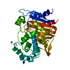


 PDBj
PDBj
 Assembly
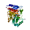

 Mass: 368.428 Da / Num. of mol.: 1 / Source method: obtained synthetically / Formula: C15H16N2O5S2
 Mass: 65.409 Da / Num. of mol.: 6 / Source method: obtained synthetically / Formula: Zn
 Mass: 22.990 Da / Num. of mol.: 1 / Source method: obtained synthetically / Formula: Na
 Mass: 18.015 Da / Num. of mol.: 373 / Source method: isolated from a natural source / Formula: H2O
 Sample preparation
 / Beamline: 31-ID / Wavelength: 0.9791 Å
 Processing