 Movie
Movie Controller

[English] 日本語
 Yorodumi
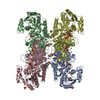
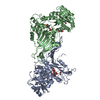
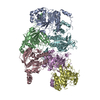
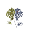
- PDB-5aq1: Trypanosoma cruzi Glucose-6-phosphate Dehydrogenase in complex wi... -
+ Open data

- Basic information
| Entry | Database: PDB / ID: 5aq1 | ||||||
|---|---|---|---|---|---|---|---|
| Title | Trypanosoma cruzi Glucose-6-phosphate Dehydrogenase in complex with G6P and NADPH | ||||||
 Components Components | GLUCOSE-6-PHOSPHATE DEHYDROGENASE | ||||||
 Keywords Keywords | OXIDOREDUCTASE / PENTOSE PHOSPHATE PATHWAY / REDOX BALANCE / CHAGAS DISEASE / G6PD / G6PDH. | ||||||
| Function / homology |  Function and homology information Function and homology informationglucose-6-phosphate dehydrogenase (NADP+) / glucose-6-phosphate dehydrogenase activity / pentose-phosphate shunt, oxidative branch / glucose metabolic process / NADP binding Similarity search - Function | ||||||
| Biological species |  | ||||||
| Method |  X-RAY DIFFRACTION / SYNCHROTRON / MOLECULAR REPLACEMENT / Resolution: 2.65 Å X-RAY DIFFRACTION / SYNCHROTRON / MOLECULAR REPLACEMENT / Resolution: 2.65 Å | ||||||
 Authors Authors | Mercaldi, G.F. / Dawson, A. / Hunter, W.N. / Cordeiro, A.T. | ||||||
 Citation Citation | Journal: FEBS Lett. / Year: 2016 Title: The Structure of a Trypanosoma Cruzi Glucose-6-Phosphate Dehydrogenase Reveals Differences from the Mammalian Enzyme. Authors: Mercaldi, G.F. / Dawson, A. / Hunter, W.N. / Cordeiro, A.T. | ||||||
| History |
|
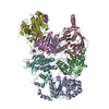
- Structure visualization
| Structure viewer | Molecule: MolmilJmol/JSmol |
|---|
- Downloads & links
-Download
| PDBx/mmCIF format | 5aq1.cif.gz | 581.7 KB | Display | PDBx/mmCIF format |
|---|---|---|---|---|
| PDB format | pdb5aq1.ent.gz | 484.8 KB | Display | PDB format |
| PDBx/mmJSON format | 5aq1.json.gz | Tree view | PDBx/mmJSON format | |
| Others |  Other downloads Other downloads |
-Validation report
| Arichive directory | https://data.pdbj.org/pub/pdb/validation_reports/aq/5aq1ftp://data.pdbj.org/pub/pdb/validation_reports/aq/5aq1 | HTTPS FTP |
|---|
-Related structure data
| Related structure data |  4e9i S: Starting model for refinement |
|---|---|
| Similar structure data |

-Links
 PDBj
PDBj

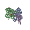
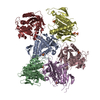
- Assembly
| Deposited unit | 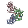
| ||||||||
|---|---|---|---|---|---|---|---|---|---|
| 1 | 
| ||||||||
| 2 | 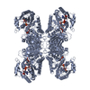
| ||||||||
| Unit cell |
|
-Components
| #1: Protein | Mass: 57620.449 Da / Num. of mol.: 3 / Fragment: UNP RESIDUES 58-545 Source method: isolated from a genetically manipulated source Source: (gene. exp.)  References: UniProt: Q4E0B2, glucose-6-phosphate dehydrogenase (NADP+) #2: Sugar | 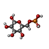  Type: D-saccharide, beta linking / Mass: 260.136 Da / Num. of mol.: 3 Type: D-saccharide, beta linking / Mass: 260.136 Da / Num. of mol.: 3Source method: isolated from a genetically manipulated source Formula: C6H13O9P #3: Chemical |   Mass: 745.421 Da / Num. of mol.: 3 / Source method: obtained synthetically / Formula: C21H30N7O17P3 Mass: 745.421 Da / Num. of mol.: 3 / Source method: obtained synthetically / Formula: C21H30N7O17P3#4: Water | ChemComp-HOH / |  Mass: 18.015 Da / Num. of mol.: 253 / Source method: isolated from a natural source / Formula: H2O Mass: 18.015 Da / Num. of mol.: 253 / Source method: isolated from a natural source / Formula: H2OHas protein modification | Y | Sequence details | TRUNCATED CONSTRUCT WITH 57 AMINOACIDS | |
|---|
-Experimental details
-Experiment
| Experiment | Method: X-RAY DIFFRACTION / Number of used crystals: 1 |
|---|
- Sample preparation
| Crystal | Density Matthews: 3.03 Å3/Da / Density % sol: 59.35 % / Description: NONE |
|---|---|
| Crystal grow | Details: PROTEIN: 2UL TCG6PD 10MG/ML, 5MM G6P, 2 MM NADPH; BUFFER: 20 MM TRIS PH 8.0 WITH 0.2 M NACL AND 5 MM ME PRECIPITANT: 1 UL JEFFAMINE ED-2003 PH 7.0 30%, 0.1 M HEPES PH 7.0; |
-Data collection
| Diffraction | Mean temperature: 100 K |
|---|---|
| Diffraction source | Source: SYNCHROTRON / Site: Diamond  / Beamline: I04-1 / Wavelength: 0.91741 / Beamline: I04-1 / Wavelength: 0.91741 |
| Detector | Type: DECTRIS PILATUS 6M / Detector: PIXEL / Date: Mar 4, 2015 |
| Radiation | Protocol: SINGLE WAVELENGTH / Monochromatic (M) / Laue (L): M / Scattering type: x-ray |
| Radiation wavelength | Wavelength: 0.91741 Å / Relative weight: 1 |
| Reflection | Resolution: 2.65→49.13 Å / Num. obs: 58510 / % possible obs: 95.4 % / Observed criterion σ(I): 2.5 / Redundancy: 8.7 % / Biso Wilson estimate: 39.6 Å2 / Rmerge(I) obs: 0.12 / Net I/σ(I): 11.1 |
| Reflection shell | Resolution: 2.65→2.72 Å / Redundancy: 8.6 % / Rmerge(I) obs: 0.79 / Mean I/σ(I) obs: 2.5 / % possible all: 96.9 |
- Processing
| Software |
| ||||||||||||||||||||||||||||||||||||||||||||||||||||||||||||||||||||||||||||||||||||||||||||||||||||||||||||||||||||||||||||||||||||||||||||||||||||||||||||||||||||||||||||||||||||||
|---|---|---|---|---|---|---|---|---|---|---|---|---|---|---|---|---|---|---|---|---|---|---|---|---|---|---|---|---|---|---|---|---|---|---|---|---|---|---|---|---|---|---|---|---|---|---|---|---|---|---|---|---|---|---|---|---|---|---|---|---|---|---|---|---|---|---|---|---|---|---|---|---|---|---|---|---|---|---|---|---|---|---|---|---|---|---|---|---|---|---|---|---|---|---|---|---|---|---|---|---|---|---|---|---|---|---|---|---|---|---|---|---|---|---|---|---|---|---|---|---|---|---|---|---|---|---|---|---|---|---|---|---|---|---|---|---|---|---|---|---|---|---|---|---|---|---|---|---|---|---|---|---|---|---|---|---|---|---|---|---|---|---|---|---|---|---|---|---|---|---|---|---|---|---|---|---|---|---|---|---|---|---|---|
| Refinement | Method to determine structure: MOLECULAR REPLACEMENT Starting model: PDB ENTRY 4E9I 4e9i Resolution: 2.65→141.57 Å / Cor.coef. Fo:Fc: 0.96 / Cor.coef. Fo:Fc free: 0.946 / SU B: 20.601 / SU ML: 0.206 / Cross valid method: THROUGHOUT / ESU R: 0.694 / ESU R Free: 0.288 / Stereochemistry target values: MAXIMUM LIKELIHOOD Details: HYDROGENS HAVE BEEN ADDED IN THE RIDING POSITIONS. U VALUES WITH TLS ADDED
| ||||||||||||||||||||||||||||||||||||||||||||||||||||||||||||||||||||||||||||||||||||||||||||||||||||||||||||||||||||||||||||||||||||||||||||||||||||||||||||||||||||||||||||||||||||||
| Solvent computation | Ion probe radii: 0.9 Å / Shrinkage radii: 0.9 Å / VDW probe radii: 1.1 Å / Solvent model: MASK | ||||||||||||||||||||||||||||||||||||||||||||||||||||||||||||||||||||||||||||||||||||||||||||||||||||||||||||||||||||||||||||||||||||||||||||||||||||||||||||||||||||||||||||||||||||||
| Displacement parameters | Biso mean: 47.798 Å2
| ||||||||||||||||||||||||||||||||||||||||||||||||||||||||||||||||||||||||||||||||||||||||||||||||||||||||||||||||||||||||||||||||||||||||||||||||||||||||||||||||||||||||||||||||||||||
| Refinement step | Cycle: LAST / Resolution: 2.65→141.57 Å
| ||||||||||||||||||||||||||||||||||||||||||||||||||||||||||||||||||||||||||||||||||||||||||||||||||||||||||||||||||||||||||||||||||||||||||||||||||||||||||||||||||||||||||||||||||||||
| Refine LS restraints |
|