 Movie
Movie Controller

[English] 日本語
 Yorodumi
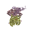
- PDB-2bhl: X-RAY STRUCTURE OF HUMAN GLUCOSE-6-PHOSPHATE DEHYDROGENASE (DELET... -
+ Open data

- Basic information
| Entry | Database: PDB / ID: 2bhl | ||||||
|---|---|---|---|---|---|---|---|
| Title | X-RAY STRUCTURE OF HUMAN GLUCOSE-6-PHOSPHATE DEHYDROGENASE (DELETION VARIANT) COMPLEXED WITH GLUCOSE-6-PHOSPHATE | ||||||
 Components Components | GLUCOSE-6-PHOSPHATE 1-DEHYDROGENASE | ||||||
 Keywords Keywords | OXIDOREDUCTASE / OXIDOREDUCTASE (CHOH(D)-NADP) / GLUCOSE METABOLISM | ||||||
| Function / homology |  Function and homology information Function and homology informationpentose biosynthetic process / ribose phosphate biosynthetic process / response to iron(III) ion / positive regulation of calcium ion transmembrane transport via high voltage-gated calcium channel / glucose-6-phosphate dehydrogenase (NADP+) / glucose-6-phosphate dehydrogenase activity / Pentose phosphate pathway / pentose-phosphate shunt, oxidative branch / negative regulation of cell growth involved in cardiac muscle cell development / glucose 6-phosphate metabolic process ...pentose biosynthetic process / ribose phosphate biosynthetic process / response to iron(III) ion / positive regulation of calcium ion transmembrane transport via high voltage-gated calcium channel / glucose-6-phosphate dehydrogenase (NADP+) / glucose-6-phosphate dehydrogenase activity / Pentose phosphate pathway / pentose-phosphate shunt, oxidative branch / negative regulation of cell growth involved in cardiac muscle cell development / glucose 6-phosphate metabolic process / NADP+ metabolic process / pentose-phosphate shunt / D-glucose binding / NFE2L2 regulates pentose phosphate pathway genes / Oxidoreductases / erythrocyte maturation / cholesterol biosynthetic process / response to food / negative regulation of reactive oxygen species metabolic process / substantia nigra development / regulation of neuron apoptotic process / TP53 Regulates Metabolic Genes / lipid metabolic process / glutathione metabolic process / glucose metabolic process / cytoplasmic side of plasma membrane / centriolar satellite / NADP binding / cellular response to oxidative stress / response to ethanol / protein homodimerization activity / extracellular exosome / membrane / identical protein binding / cytoplasm / cytosol Similarity search - Function | ||||||
| Biological species |  HOMO SAPIENS (human) HOMO SAPIENS (human) | ||||||
| Method |  X-RAY DIFFRACTION / SYNCHROTRON / MOLECULAR REPLACEMENT / Resolution: 2.9 Å X-RAY DIFFRACTION / SYNCHROTRON / MOLECULAR REPLACEMENT / Resolution: 2.9 Å | ||||||
 Authors Authors | Kotaka, M. / Gover, S. / Lam, V.M.S. / Adams, M.J. | ||||||
 Citation Citation | Journal: Acta Crystallogr.,Sect.D / Year: 2005 Title: Structural Studies of Glucose-6-Phosphate and Nadp+ Binding to Human Glucose-6-Phosphate Dehydrogenase Authors: Kotaka, M. / Gover, S. / Vandeputte-Rutten, L. / Au, S.W.N. / Lam, V.M.S. / Adams, M.J. #1: Journal: Structure / Year: 2000Title: Human Glucose-6-Phosphate Dehydrogenase: The Crystal Structure Reveals a Structural Nadp Molecule and Provides Insights Into Enzyme Deficiency Authors: Au, S.W.N. / Gover, S. / Lam, V.M.S. / Adams, M.J. #2: Journal: Acta Crystallogr.,Sect.D / Year: 1999 Title: Solution of the Structure of Tetrameric Human Glucose 6-Phosphate Dehydrogenase by Molecular Replacement Authors: Au, S.W.N. / Naylor, C.E. / Gover, S. / Vandeputte-Rutten, L. / Scopes, D.A. / Mason, P.J. / Luzzatto, L. / Lam, V.M.S. / Adams, M.J. | ||||||
| History |
| ||||||
| Remark 650 | HELIX DETERMINATION METHOD: PROCHECK, WITH IDENTIFICATION CORRESPONDING TO THE 2.0A LEUCONOSTOC ... HELIX DETERMINATION METHOD: PROCHECK, WITH IDENTIFICATION CORRESPONDING TO THE 2.0A LEUCONOSTOC MESENTERODES STRUCTURE, 1DPG. THE BEND AT LYS 47 IN HELIX A IS A CONSEQUENCE OF THE CONSERVED PRO 50. | ||||||
| Remark 700 | SHEET DETERMINATION METHOD: PROCHECK WITH EXTENSION TAKEN WHERE HYDROGEN BONDING INDICATES THAT ... SHEET DETERMINATION METHOD: PROCHECK WITH EXTENSION TAKEN WHERE HYDROGEN BONDING INDICATES THAT THIS IS APPROPRIATE |
- Structure visualization
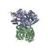
| Structure viewer | Molecule: MolmilJmol/JSmol |
|---|
- Downloads & links
-Download
| PDBx/mmCIF format | 2bhl.cif.gz | 204.7 KB | Display | PDBx/mmCIF format |
|---|---|---|---|---|
| PDB format | pdb2bhl.ent.gz | 163.8 KB | Display | PDB format |
| PDBx/mmJSON format | 2bhl.json.gz | Tree view | PDBx/mmJSON format | |
| Others |  Other downloads Other downloads |
-Validation report
| Arichive directory | https://data.pdbj.org/pub/pdb/validation_reports/bh/2bhlftp://data.pdbj.org/pub/pdb/validation_reports/bh/2bhl | HTTPS FTP |
|---|
-Related structure data
| Related structure data | 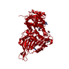 2bh9C 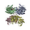 1qkiS S: Starting model for refinement C: citing same article ( |
|---|---|
| Similar structure data |


-Links
 PDBj
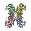
PDBj- Assembly
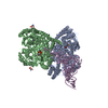
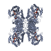
| Deposited unit | 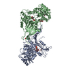
| ||||||||
|---|---|---|---|---|---|---|---|---|---|
| 1 | 
| ||||||||
| Unit cell |
| ||||||||
| Noncrystallographic symmetry (NCS) | NCS oper: (Code: given Matrix: (-0.99911, -0.00678, -0.04166), Vector: |
-Components
| #1: Protein | Mass: 56397.258 Da / Num. of mol.: 2 / Mutation: YES Source method: isolated from a genetically manipulated source Details: THE 25 N-TERMINAL RESIDUES HAVE BEEN REMOVED AND THE FIRST RESIDUE IS VALINE, NOT HISTIDINE Source: (gene. exp.) HOMO SAPIENS (human) / Plasmid: PTRC99A/DG6PD / Production host:  References: UniProt: P11413, glucose-6-phosphate dehydrogenase (NADP+) #2: Chemical | ChemComp-GOL /   Mass: 92.094 Da / Num. of mol.: 6 / Source method: obtained synthetically / Formula: C3H8O3 Mass: 92.094 Da / Num. of mol.: 6 / Source method: obtained synthetically / Formula: C3H8O3#3: Sugar | 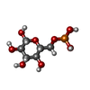  Type: D-saccharide, beta linking / Mass: 260.136 Da / Num. of mol.: 2 Type: D-saccharide, beta linking / Mass: 260.136 Da / Num. of mol.: 2Source method: isolated from a genetically manipulated source Formula: C6H13O9P #4: Water | ChemComp-HOH / |  Mass: 18.015 Da / Num. of mol.: 81 / Source method: isolated from a natural source / Formula: H2O Mass: 18.015 Da / Num. of mol.: 81 / Source method: isolated from a natural source / Formula: H2O |
|---|
-Experimental details
-Experiment
| Experiment | Method: X-RAY DIFFRACTION / Number of used crystals: 1 |
|---|
- Sample preparation
| Crystal | Density Matthews: 3.1 Å3/Da / Density % sol: 59.8 % |
|---|---|
| Crystal grow | pH: 8.5 Details: 0.1M TRIS, PH 8.5; 0.2M MGCL2; 12% PEG 4000; 5% GLYCEROL |
-Data collection
| Diffraction | Mean temperature: 100 K |
|---|---|
| Diffraction source | Source: SYNCHROTRON / Site: SRS  / Beamline: PX14.1 / Wavelength: 1.488 / Beamline: PX14.1 / Wavelength: 1.488 |
| Detector | Type: ADSC CCD / Detector: CCD / Date: Nov 27, 2002 / Details: MIRRORS |
| Radiation | Monochromator: SI CRYSTAL / Protocol: SINGLE WAVELENGTH / Monochromatic (M) / Laue (L): M / Scattering type: x-ray |
| Radiation wavelength | Wavelength: 1.488 Å / Relative weight: 1 |
| Reflection | Resolution: 2.9→69 Å / Num. obs: 32261 / % possible obs: 98.9 % / Observed criterion σ(I): -4 / Redundancy: 6.8 % / Rmerge(I) obs: 0.12 / Net I/σ(I): 5.3 |
| Reflection shell | Resolution: 2.9→3.07 Å / Redundancy: 2.2 % / Rmerge(I) obs: 0.41 / Mean I/σ(I) obs: 2 / % possible all: 92.9 |
- Processing
| Software |
| |||||||||||||||||||||||||||||||||||||||||||||||||||||||||
|---|---|---|---|---|---|---|---|---|---|---|---|---|---|---|---|---|---|---|---|---|---|---|---|---|---|---|---|---|---|---|---|---|---|---|---|---|---|---|---|---|---|---|---|---|---|---|---|---|---|---|---|---|---|---|---|---|---|---|
| Refinement | Method to determine structure: MOLECULAR REPLACEMENT Starting model: PDB ENTRY 1QKI CHAIN A, B Resolution: 2.9→69 Å / Cross valid method: FREE R-VALUE / σ(F): 0 Details: RESIDUES 506 TO 515 WERE DISORDERED AND NOT INCLUDED
| |||||||||||||||||||||||||||||||||||||||||||||||||||||||||
| Refinement step | Cycle: LAST / Resolution: 2.9→69 Å
| |||||||||||||||||||||||||||||||||||||||||||||||||||||||||
| Refine LS restraints |
|
