 Movie
Movie Controller

[English] 日本語
 Yorodumi
- PDB-2bh9: X-RAY STRUCTURE OF A DELETION VARIANT OF HUMAN GLUCOSE 6-PHOSPHAT... -
+ Open data

- Basic information
| Entry | Database: PDB / ID: 2bh9 | ||||||
|---|---|---|---|---|---|---|---|
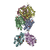
| Title | X-RAY STRUCTURE OF A DELETION VARIANT OF HUMAN GLUCOSE 6-PHOSPHATE DEHYDROGENASE COMPLEXED WITH STRUCTURAL AND COENZYME NADP | ||||||
 Components Components | GLUCOSE-6-PHOSPHATE 1-DEHYDROGENASE | ||||||
 Keywords Keywords | OXIDOREDUCTASE / OXIDOREDUCTASE (CHOH(D)-NADP) / CARBOHYDRATE METABOLISM / GLUCOSE METABOLISM | ||||||
| Function / homology |  Function and homology information Function and homology informationpentose biosynthetic process / ribose phosphate biosynthetic process / response to iron(III) ion / positive regulation of calcium ion transmembrane transport via high voltage-gated calcium channel / glucose-6-phosphate dehydrogenase (NADP+) / glucose-6-phosphate dehydrogenase activity / Pentose phosphate pathway / pentose-phosphate shunt, oxidative branch / negative regulation of cell growth involved in cardiac muscle cell development / glucose 6-phosphate metabolic process ...pentose biosynthetic process / ribose phosphate biosynthetic process / response to iron(III) ion / positive regulation of calcium ion transmembrane transport via high voltage-gated calcium channel / glucose-6-phosphate dehydrogenase (NADP+) / glucose-6-phosphate dehydrogenase activity / Pentose phosphate pathway / pentose-phosphate shunt, oxidative branch / negative regulation of cell growth involved in cardiac muscle cell development / glucose 6-phosphate metabolic process / NADP+ metabolic process / pentose-phosphate shunt / D-glucose binding / NFE2L2 regulates pentose phosphate pathway genes / Oxidoreductases / cholesterol biosynthetic process / erythrocyte maturation / response to food / negative regulation of reactive oxygen species metabolic process / substantia nigra development / regulation of neuron apoptotic process / TP53 Regulates Metabolic Genes / lipid metabolic process / glutathione metabolic process / glucose metabolic process / cytoplasmic side of plasma membrane / centriolar satellite / NADP binding / cellular response to oxidative stress / response to ethanol / protein homodimerization activity / extracellular exosome / membrane / identical protein binding / cytoplasm / cytosol Similarity search - Function | ||||||
| Biological species |  HOMO SAPIENS (human) HOMO SAPIENS (human) | ||||||
| Method |  X-RAY DIFFRACTION / SYNCHROTRON / MOLECULAR REPLACEMENT / Resolution: 2.5 Å X-RAY DIFFRACTION / SYNCHROTRON / MOLECULAR REPLACEMENT / Resolution: 2.5 Å | ||||||
 Authors Authors | Gover, S. / Vandeputte-Rutten, L. / Au, S.W.N. / Adams, M.J. | ||||||
 Citation Citation | Journal: Acta Crystallogr.,Sect.D / Year: 2005 Title: Structural Studies of Glucose-6-Phosphate and Nadp+ Binding to Human Glucose-6-Phosphate Dehydrogenase Authors: Kotaka, M. / Gover, S. / Vandeputte-Rutten, L. / Au, S.W.N. / Lam, V.M.S. / Adams, M.J. #1: Journal: Structure / Year: 2000Title: Human Glucose-6-Phosphate Dehydrogenase: The Crystal Structure Reveals a Structural Nadp Molecule and Provides Insights Into Enzyme Deficiency Authors: Au, S.W.N. / Gover, S. / Lam, V.M.S. / Adams, M.J. #2: Journal: Acta Crystallogr.,Sect.D / Year: 1999 Title: Solution of the Structure of Tetrameric Human Glucose 6-Phosphate Dehydrogenase by Molecular Replacement Authors: Au, S.W.N. / Naylor, C.E. / Gover, S. / Vandeputte-Rutten, L. / Scopes, D.A. / Mason, P.J. / Luzzatto, L. / Lam, V.M.S. / Adams, M.J. | ||||||
| History |
|
- Structure visualization
| Structure viewer | Molecule: MolmilJmol/JSmol |
|---|
- Downloads & links
-Download
| PDBx/mmCIF format | 2bh9.cif.gz | 117.4 KB | Display | PDBx/mmCIF format |
|---|---|---|---|---|
| PDB format | pdb2bh9.ent.gz | 90.4 KB | Display | PDB format |
| PDBx/mmJSON format | 2bh9.json.gz | Tree view | PDBx/mmJSON format | |
| Others |  Other downloads Other downloads |
-Validation report
| Arichive directory | https://data.pdbj.org/pub/pdb/validation_reports/bh/2bh9ftp://data.pdbj.org/pub/pdb/validation_reports/bh/2bh9 | HTTPS FTP |
|---|
-Related structure data


-Links
 PDBj
PDBj- Assembly
| Deposited unit | 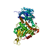
| ||||||||||||||||||||||||
|---|---|---|---|---|---|---|---|---|---|---|---|---|---|---|---|---|---|---|---|---|---|---|---|---|---|
| 1 | 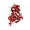
| ||||||||||||||||||||||||
| Unit cell |
| ||||||||||||||||||||||||
| Components on special symmetry positions |
|
-Components
| #1: Protein | Mass: 56397.258 Da / Num. of mol.: 1 / Fragment: RESIDUES 26-514 Source method: isolated from a genetically manipulated source Source: (gene. exp.) HOMO SAPIENS (human) / Plasmid: PKKG6PD / Production host:  References: UniProt: P11413, glucose-6-phosphate dehydrogenase (NADP+) | ||||||||
|---|---|---|---|---|---|---|---|---|---|
| #2: Chemical |   Mass: 743.405 Da / Num. of mol.: 2 / Source method: obtained synthetically / Formula: C21H28N7O17P3 Mass: 743.405 Da / Num. of mol.: 2 / Source method: obtained synthetically / Formula: C21H28N7O17P3#3: Chemical |   Mass: 92.094 Da / Num. of mol.: 2 / Source method: obtained synthetically / Formula: C3H8O3 Mass: 92.094 Da / Num. of mol.: 2 / Source method: obtained synthetically / Formula: C3H8O3#4: Water | ChemComp-HOH / |  Mass: 18.015 Da / Num. of mol.: 162 / Source method: isolated from a natural source / Formula: H2O Mass: 18.015 Da / Num. of mol.: 162 / Source method: isolated from a natural source / Formula: H2OCompound details | ENGINEERED | Sequence details | THE G6PD USED IN THIS WORK IS AN ENGINEERED VARIANT IN WHICH THE 25 N-TERMINAL RESIDUES HAVE BEEN ...THE G6PD USED IN THIS WORK IS AN ENGINEERED | |
-Experimental details
-Experiment
| Experiment | Method: X-RAY DIFFRACTION / Number of used crystals: 1 |
|---|
- Sample preparation
| Crystal | Density Matthews: 2.53 Å3/Da / Density % sol: 51.4 % |
|---|---|
| Crystal grow | Method: vapor diffusion, hanging drop / pH: 8 Details: HANGING DROP VAPOUR DIFFUSION. 1-PLUS-1 MICROLITRE DROPS IN THE WELL 0.1M TRIS-HCL,PH 7.5-8.2, 10-16% PEG 4000. PROTEIN CONCENTRATION 5MG/ML. |
-Data collection
| Diffraction | Mean temperature: 100 K |
|---|---|
| Diffraction source | Source: SYNCHROTRON / Site: SRS  / Beamline: PX9.6 / Wavelength: 0.87 / Beamline: PX9.6 / Wavelength: 0.87 |
| Detector | Type: MARRESEARCH / Detector: IMAGE PLATE / Date: Oct 21, 1996 / Details: MIRRORS |
| Radiation | Monochromator: SI CRYSTAL / Protocol: SINGLE WAVELENGTH / Monochromatic (M) / Laue (L): M / Scattering type: x-ray |
| Radiation wavelength | Wavelength: 0.87 Å / Relative weight: 1 |
| Reflection | Resolution: 2.5→29.7 Å / Num. obs: 14581 / % possible obs: 73.3 % / Observed criterion σ(I): -4 / Redundancy: 2.5 % / Rmerge(I) obs: 0.12 / Net I/σ(I): 9.6 |
| Reflection shell | Resolution: 2.5→2.64 Å / Redundancy: 2.2 % / Rmerge(I) obs: 0.31 / Mean I/σ(I) obs: 3.4 / % possible all: 59.3 |
- Processing
| Software |
| ||||||||||||||||||||
|---|---|---|---|---|---|---|---|---|---|---|---|---|---|---|---|---|---|---|---|---|---|
| Refinement | Method to determine structure: MOLECULAR REPLACEMENT Starting model: PARTIALLY-REFINED MONOMER OF HUMAN G6PD, CANTON VARIANT Resolution: 2.5→29.7 Å / Cross valid method: FREE THROUGHOUT / σ(F): 0
| ||||||||||||||||||||
| Refinement step | Cycle: LAST / Resolution: 2.5→29.7 Å
|
