[English] 日本語
Yorodumi
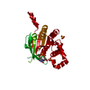
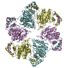
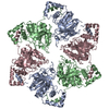
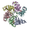
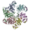
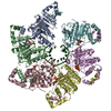
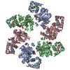
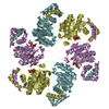
- PDB-1e0j: gp4d helicase from phage T7 ADPNP complex -

+
Open data


ID or keywords:

Loading...

-
Basic information

Entry
Database: PDB / ID: 1e0j
Titlegp4d helicase from phage T7 ADPNP complex
ComponentsDNA HELICASEHelicase
KeywordsHELICASE / ATPASE / DNA REPLICATION
Function / homology
Function and homology information


DNA primase activity / 5'-3' DNA helicase activity / viral DNA genome replication / DNA helicase activity / Transferases; Transferring phosphorus-containing groups; Nucleotidyltransferases / single-stranded DNA binding / DNA helicase / ATP hydrolysis activity / zinc ion binding / ATP binding / identical protein binding
Similarity search - Function
Bacteriophage T7 DNA helicase/primase / : / Bacteriophage T7 DNA helicase/primase, N-terminal a+b fold / Bacteriophage T7, Gp4, DNA primase/helicase, N-terminal / Zinc-binding domain of primase-helicase / Zinc-binding domain of primase-helicase / Twinkle-like protein / Archaeal primase DnaG/twinkle-like, TOPRIM domain / Toprim-like / DnaB-like helicase C terminal domain ...Bacteriophage T7 DNA helicase/primase / : / Bacteriophage T7 DNA helicase/primase, N-terminal a+b fold / Bacteriophage T7, Gp4, DNA primase/helicase, N-terminal / Zinc-binding domain of primase-helicase / Zinc-binding domain of primase-helicase / Twinkle-like protein / Archaeal primase DnaG/twinkle-like, TOPRIM domain / Toprim-like / DnaB-like helicase C terminal domain / DNA helicase, DnaB-like, C-terminal / Superfamily 4 helicase domain profile. / TOPRIM / Toprim domain profile. / TOPRIM domain / P-loop containing nucleotide triphosphate hydrolases / P-loop containing nucleoside triphosphate hydrolase / Rossmann fold / 3-Layer(aba) Sandwich / Alpha Beta
Similarity search - Domain/homology
PHOSPHOAMINOPHOSPHONIC ACID-ADENYLATE ESTER / DNA helicase/primase
Similarity search - Component
Biological speciesPHAGE T7 (virus)
MethodX-RAY DIFFRACTION / SYNCHROTRON / MOLECULAR REPLACEMENT / Resolution: 3 Å
AuthorsSingleton, M.R. / Sawaya, M.R. / Ellenberger, T. / Wigley, D.B.
CitationJournal: Cell(Cambridge,Mass.) / Year: 2000
Title: Crystal Structure of T7 Gene 4 Ring Helicase Indicates a Mechanism for Sequential Hydrolysis of Nucleotides
Authors: Singleton, M.R. / Sawaya, M.R. / Ellenberger, T. / Wigley, D.B.
History
DepositionMar 30, 2000Deposition site: PDBE / Processing site: PDBE
Revision 1.0Jun 9, 2000Provider: repository / Type: Initial release
Revision 1.1May 8, 2011Group: Version format compliance
Revision 1.2Jul 13, 2011Group: Version format compliance

-
Structure visualization

Structure viewerMolecule:
MolmilJmol/JSmol

Downloads & links

-
Assembly

Deposited unit
A: DNA HELICASE
B: DNA HELICASE
C: DNA HELICASE
D: DNA HELICASE
E: DNA HELICASE
F: DNA HELICASE
hetero molecules


Theoretical massNumber of molelcules
Total (without water)192,65314
Polymers190,5316
Non-polymers2,1228
Water0
1
A: DNA HELICASE
B: DNA HELICASE
C: DNA HELICASE
hetero molecules

A: DNA HELICASE
B: DNA HELICASE
C: DNA HELICASE
hetero molecules


Theoretical massNumber of molelcules
Total (without water)192,65314
Polymers190,5316
Non-polymers2,1228
Water0
TypeNameSymmetry operationNumber
identity operation1_555x,y,z1
crystal symmetry operation7_556y,x,-z+11
MethodPQS
2
D: DNA HELICASE
E: DNA HELICASE
F: DNA HELICASE
hetero molecules

D: DNA HELICASE
E: DNA HELICASE
F: DNA HELICASE
hetero molecules


Theoretical massNumber of molelcules
Total (without water)192,65314
Polymers190,5316
Non-polymers2,1228
Water0
TypeNameSymmetry operationNumber
identity operation1_555x,y,z1
crystal symmetry operation7_556y,x,-z+11
MethodPQS
Unit cell
Length a, b, c (Å)119.180, 119.180, 283.420
Angle α, β, γ (deg.)90.00, 90.00, 90.00
Int Tables number92
Space group name H-MP41212

-
Components

#1: Protein
DNA HELICASE / Helicase


Mass: 31755.139 Da / Num. of mol.: 6 / Fragment: DOMAIN 4D
Source method: isolated from a genetically manipulated source
Source: (gene. exp.) PHAGE T7 (virus) / Gene: GENE 4 / Production host: ESCHERICHIA COLI (E. coli) / References: UniProt: P03692
#2: Chemical
ChemComp-ANP / PHOSPHOAMINOPHOSPHONIC ACID-ADENYLATE ESTER


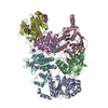
Mass: 506.196 Da / Num. of mol.: 4 / Source method: obtained synthetically / Formula: C10H17N6O12P3 / Comment: AMP-PNP, energy-carrying molecule analogue*YM
#3: Chemical
ChemComp-MG / MAGNESIUM ION


Mass: 24.305 Da / Num. of mol.: 4 / Source method: obtained synthetically / Formula: Mg

-
Experimental details

-
Experiment

ExperimentMethod: X-RAY DIFFRACTION / Number of used crystals: 1

-
Sample preparation

CrystalDensity Matthews: 2.64 Å3/Da / Density % sol: 53.43 %
Crystal growpH: 9.5 / Details: pH 9.50

-
Data collection

DiffractionMean temperature: 100 K
Diffraction sourceSource: SYNCHROTRON / Site: SRS / Beamline: PX7.2 / Wavelength: 1.488
DetectorType: MARRESEARCH / Detector: IMAGE PLATE
RadiationProtocol: SINGLE WAVELENGTH / Monochromatic (M) / Laue (L): M / Scattering type: x-ray
Radiation wavelengthWavelength: 1.488 Å / Relative weight: 1
ReflectionResolution: 3→20 Å / Num. obs: 40673 / % possible obs: 97.9 % / Redundancy: 3 % / Rsym value: 0.065 / Net I/σ(I): 16.5
Reflection shellResolution: 3→3.11 Å / Redundancy: 3 % / Rsym value: 0.304 / % possible all: 98.9

-
Processing

Software
NameVersionClassification
CNS1refinement
DENZOdata reduction
SCALEPACKdata scaling
CNS1phasing
RefinementMethod to determine structure: MOLECULAR REPLACEMENT / Resolution: 3→20 Å / Rfactor Rfree error: 0.007 / Cross valid method: THROUGHOUT / σ(F): 0
RfactorNum. reflection% reflectionSelection details
Rfree0.304 2107 5.2 %RANDOM
Rwork0.253 ---
obs0.253 40644 96.7 %-
Refinement stepCycle: LAST / Resolution: 3→20 Å
ProteinNucleic acidLigandSolventTotal
Num. atoms13264 0 128 0 13392
Refine LS restraints
Refine-IDTypeDev ideal
X-RAY DIFFRACTIONc_bond_d0.009
X-RAY DIFFRACTIONc_bond_d_na
X-RAY DIFFRACTIONc_bond_d_prot
X-RAY DIFFRACTIONc_angle_d
X-RAY DIFFRACTIONc_angle_d_na
X-RAY DIFFRACTIONc_angle_d_prot
X-RAY DIFFRACTIONc_angle_deg1.5
X-RAY DIFFRACTIONc_angle_deg_na
X-RAY DIFFRACTIONc_angle_deg_prot
X-RAY DIFFRACTIONc_dihedral_angle_d24
X-RAY DIFFRACTIONc_dihedral_angle_d_na
X-RAY DIFFRACTIONc_dihedral_angle_d_prot
X-RAY DIFFRACTIONc_improper_angle_d0.8
X-RAY DIFFRACTIONc_improper_angle_d_na
X-RAY DIFFRACTIONc_improper_angle_d_prot
X-RAY DIFFRACTIONc_mcbond_it
X-RAY DIFFRACTIONc_mcangle_it
X-RAY DIFFRACTIONc_scbond_it
X-RAY DIFFRACTIONc_scangle_it
Refine LS restraints NCSNCS model details: RESTRAINTS
LS refinement shellResolution: 3→3.19 Å / Rfactor Rfree error: 0.021 / Total num. of bins used: 6
RfactorNum. reflection% reflection
Rfree0.406 379 5.8 %
Rwork0.37 6196 -
obs--96.7 %

+
About Yorodumi

-
News

-
Feb 9, 2022. New format data for meta-information of EMDB entries

New format data for meta-information of EMDB entries

  • Version 3 of the EMDB header file is now the official format.
  • The previous official version 1.9 will be removed from the archive.

Related info.:EMDB header

External links:wwPDB to switch to version 3 of the EMDB data model

-
Aug 12, 2020. Covid-19 info

Covid-19 info

URL: https://pdbj.org/emnavi/covid19.php

New page: Covid-19 featured information page in EM Navigator.

Related info.:Covid-19 info / Mar 5, 2020. Novel coronavirus structure data

+
Mar 5, 2020. Novel coronavirus structure data

Novel coronavirus structure data

Related info.:Yorodumi Speices / Aug 12, 2020. Covid-19 info

External links:COVID-19 featured content - PDBj / Molecule of the Month (242):Coronavirus Proteases

+
Jan 31, 2019. EMDB accession codes are about to change! (news from PDBe EMDB page)

EMDB accession codes are about to change! (news from PDBe EMDB page)

  • The allocation of 4 digits for EMDB accession codes will soon come to an end. Whilst these codes will remain in use, new EMDB accession codes will include an additional digit and will expand incrementally as the available range of codes is exhausted. The current 4-digit format prefixed with “EMD-” (i.e. EMD-XXXX) will advance to a 5-digit format (i.e. EMD-XXXXX), and so on. It is currently estimated that the 4-digit codes will be depleted around Spring 2019, at which point the 5-digit format will come into force.
  • The EM Navigator/Yorodumi systems omit the EMD- prefix.

Related info.:Q: What is EMD? / ID/Accession-code notation in Yorodumi/EM Navigator

External links:EMDB Accession Codes are Changing Soon! / Contact to PDBj

+
Jul 12, 2017. Major update of PDB

Major update of PDB

  • wwPDB released updated PDB data conforming to the new PDBx/mmCIF dictionary.
  • This is a major update changing the version number from 4 to 5, and with Remediation, in which all the entries are updated.
  • In this update, many items about electron microscopy experimental information are reorganized (e.g. em_software).
  • Now, EM Navigator and Yorodumi are based on the updated data.

External links:wwPDB Remediation / Enriched Model Files Conforming to OneDep Data Standards Now Available in the PDB FTP Archive

-
Yorodumi

Thousand views of thousand structures

  • Yorodumi is a browser for structure data from EMDB, PDB, SASBDB, etc.
  • This page is also the successor to EM Navigator detail page, and also detail information page/front-end page for Omokage search.
  • The word "yorodu" (or yorozu) is an old Japanese word meaning "ten thousand". "mi" (miru) is to see.

Related info.:EMDB / PDB / SASBDB / Comparison of 3 databanks / Yorodumi Search / Aug 31, 2016. New EM Navigator & Yorodumi / Yorodumi Papers / Jmol/JSmol / Function and homology information / Changes in new EM Navigator and Yorodumi

Read more