 Movie
Movie Controller

[English] 日本語
 Yorodumi
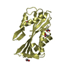
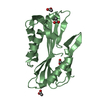
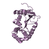
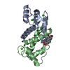
- PDB-5afp: Neuronal calcium sensor-1 (NCS-1)from Rattus norvegicus complex w... -
+ Open data

- Basic information
| Entry | Database: PDB / ID: 5afp | |||||||||
|---|---|---|---|---|---|---|---|---|---|---|
| Title | Neuronal calcium sensor-1 (NCS-1)from Rattus norvegicus complex with rhodopsin kinase peptide from Homo sapiens | |||||||||
 Components Components |
| |||||||||
 Keywords Keywords | SIGNALING PROTEIN | |||||||||
| Function / homology |  Function and homology information Function and homology informationcalcium-dependent protein kinase inhibitor activity / rhodopsin kinase / rhodopsin kinase activity / calcium sensitive guanylate cyclase activator activity / regulation of opsin-mediated signaling pathway / G protein-coupled opsin signaling pathway / regulation of presynaptic cytosolic calcium ion concentration / dense core granule / regulation of G protein-coupled receptor signaling pathway / regulation of neuron projection development ...calcium-dependent protein kinase inhibitor activity / rhodopsin kinase / rhodopsin kinase activity / calcium sensitive guanylate cyclase activator activity / regulation of opsin-mediated signaling pathway / G protein-coupled opsin signaling pathway / regulation of presynaptic cytosolic calcium ion concentration / dense core granule / regulation of G protein-coupled receptor signaling pathway / regulation of neuron projection development / regulation of synaptic vesicle exocytosis / positive regulation of exocytosis / regulation of signal transduction / postsynaptic cytosol / voltage-gated calcium channel activity / presynaptic cytosol / visual perception / positive regulation of calcium-mediated signaling / calyx of Held / photoreceptor disc membrane / protein autophosphorylation / Inactivation, recovery and regulation of the phototransduction cascade / protein kinase activity / postsynaptic density / postsynapse / axon / calcium ion binding / dendrite / protein kinase binding / perinuclear region of cytoplasm / glutamatergic synapse / magnesium ion binding / Golgi apparatus / ATP binding / plasma membrane / cytoplasm / cytosol Similarity search - Function | |||||||||
| Biological species |   HOMO SAPIENS (human) HOMO SAPIENS (human) | |||||||||
| Method |  X-RAY DIFFRACTION / SYNCHROTRON / MOLECULAR REPLACEMENT / Resolution: 2.3 Å X-RAY DIFFRACTION / SYNCHROTRON / MOLECULAR REPLACEMENT / Resolution: 2.3 Å | |||||||||
 Authors Authors | Saleem, M. / Karuppiah, V. / Pandalaneni, S. / Burgoyne, R. / Derrick, J.P. / Lian, L.Y. | |||||||||
 Citation Citation | Journal: J.Biol.Chem. / Year: 2015 Title: Neuronal Calcium Sensor-1 Binds the D2 Dopamine Receptor and G-Protein Coupled Receptor Kinase 1 (Grk1) Peptides Using Different Modes of Interactions. Authors: Pandalaneni, S. / Karuppiah, V. / Saleem, M. / Haynes, L.P. / Burgoyne, R.D. / Mayans, O. / Derrick, J.P. / Lian, L.Y. | |||||||||
| History |
|
- Structure visualization
| Structure viewer | Molecule: MolmilJmol/JSmol |
|---|
- Downloads & links
-Download
| PDBx/mmCIF format | 5afp.cif.gz | 161.7 KB | Display | PDBx/mmCIF format |
|---|---|---|---|---|
| PDB format | pdb5afp.ent.gz | 128.8 KB | Display | PDB format |
| PDBx/mmJSON format | 5afp.json.gz | Tree view | PDBx/mmJSON format | |
| Others |  Other downloads Other downloads |
-Validation report
| Arichive directory | https://data.pdbj.org/pub/pdb/validation_reports/af/5afpftp://data.pdbj.org/pub/pdb/validation_reports/af/5afp | HTTPS FTP |
|---|
-Related structure data
| Related structure data |  4yruC  5aeqC  5aerC  2you S: Starting model for refinement C: citing same article ( |
|---|---|
| Similar structure data |


-Links
 PDBj
PDBj


- Assembly
| Deposited unit | 
| ||||||||
|---|---|---|---|---|---|---|---|---|---|
| 1 | 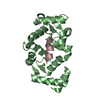
| ||||||||
| 2 | 
| ||||||||
| Unit cell |
| ||||||||
| Noncrystallographic symmetry (NCS) | NCS oper: (Code: given / Matrix: (1), |
-Components
| #1: Protein | Mass: 21902.668 Da / Num. of mol.: 2 Source method: isolated from a genetically manipulated source Source: (gene. exp.)  #2: Protein/peptide | Mass: 2550.777 Da / Num. of mol.: 2 / Fragment: RESIDUES 1-25 / Source method: obtained synthetically / Source: (synth.) HOMO SAPIENS (human) / References: UniProt: Q15835#3: Chemical | ChemComp-CA /   Mass: 40.078 Da / Num. of mol.: 6 / Source method: obtained synthetically / Formula: Ca Mass: 40.078 Da / Num. of mol.: 6 / Source method: obtained synthetically / Formula: Ca#4: Chemical |   Mass: 22.990 Da / Num. of mol.: 2 / Source method: obtained synthetically / Formula: Na Mass: 22.990 Da / Num. of mol.: 2 / Source method: obtained synthetically / Formula: Na#5: Water | ChemComp-HOH / |  Mass: 18.015 Da / Num. of mol.: 26 / Source method: isolated from a natural source / Formula: H2O Mass: 18.015 Da / Num. of mol.: 26 / Source method: isolated from a natural source / Formula: H2O |
|---|
-Experimental details
-Experiment
| Experiment | Method: X-RAY DIFFRACTION / Number of used crystals: 1 |
|---|
- Sample preparation
| Crystal | Density Matthews: 2.6 Å3/Da / Density % sol: 52 % / Description: NONE |
|---|---|
| Crystal grow | pH: 7.5 Details: 0.12M ALCOHOLS (1,6-HEXANEDIOL; 1- BUTANOL;1,PROPANEDIOL(RACEMIC); 2-PROPONOL; 1, 4-BUTANEDIOL; 1,3- PROPANEDIOL); 0.1M BUFFER 2 (SODIUM HEPES; MOPS ACID PH 7.5); P550MME_P20K 30%. |
-Data collection
| Diffraction | Mean temperature: 100 K |
|---|---|
| Diffraction source | Source: SYNCHROTRON / Site: Diamond  / Beamline: I03 / Wavelength: 0.9 / Beamline: I03 / Wavelength: 0.9 |
| Detector | Date: Jul 12, 2014 |
| Radiation | Protocol: SINGLE WAVELENGTH / Monochromatic (M) / Laue (L): M / Scattering type: x-ray |
| Radiation wavelength | Wavelength: 0.9 Å / Relative weight: 1 |
| Reflection | Resolution: 2.3→48 Å / Num. obs: 18488 / % possible obs: 99.2 % / Observed criterion σ(I): 2 / Redundancy: 3.3 % / Rmerge(I) obs: 0.04 / Net I/σ(I): 15.8 |
| Reflection shell | Resolution: 2.3→2.36 Å / Redundancy: 2.6 % / Rmerge(I) obs: 0.37 / Mean I/σ(I) obs: 2.2 / % possible all: 94.3 |
- Processing
| Software |
| ||||||||||||||||||||||||||||||||||||||||||||||||||||||||||||||||||||||||||||||||||||||||||||||||||||||||||||||||||||||||||||||||||||||||||||||||||||||||||||||||||||||||||||||||||||||
|---|---|---|---|---|---|---|---|---|---|---|---|---|---|---|---|---|---|---|---|---|---|---|---|---|---|---|---|---|---|---|---|---|---|---|---|---|---|---|---|---|---|---|---|---|---|---|---|---|---|---|---|---|---|---|---|---|---|---|---|---|---|---|---|---|---|---|---|---|---|---|---|---|---|---|---|---|---|---|---|---|---|---|---|---|---|---|---|---|---|---|---|---|---|---|---|---|---|---|---|---|---|---|---|---|---|---|---|---|---|---|---|---|---|---|---|---|---|---|---|---|---|---|---|---|---|---|---|---|---|---|---|---|---|---|---|---|---|---|---|---|---|---|---|---|---|---|---|---|---|---|---|---|---|---|---|---|---|---|---|---|---|---|---|---|---|---|---|---|---|---|---|---|---|---|---|---|---|---|---|---|---|---|---|
| Refinement | Method to determine structure: MOLECULAR REPLACEMENT Starting model: PDB ENTRY 2YOU 2you Resolution: 2.3→47.86 Å / Cor.coef. Fo:Fc: 0.938 / Cor.coef. Fo:Fc free: 0.91 / SU B: 17.091 / SU ML: 0.198 / Cross valid method: THROUGHOUT / ESU R: 0.395 / ESU R Free: 0.249 / Stereochemistry target values: MAXIMUM LIKELIHOOD / Details: HYDROGENS HAVE BEEN ADDED IN THE RIDING POSITIONS.
| ||||||||||||||||||||||||||||||||||||||||||||||||||||||||||||||||||||||||||||||||||||||||||||||||||||||||||||||||||||||||||||||||||||||||||||||||||||||||||||||||||||||||||||||||||||||
| Solvent computation | Ion probe radii: 0.8 Å / Shrinkage radii: 0.8 Å / VDW probe radii: 1.2 Å / Solvent model: MASK | ||||||||||||||||||||||||||||||||||||||||||||||||||||||||||||||||||||||||||||||||||||||||||||||||||||||||||||||||||||||||||||||||||||||||||||||||||||||||||||||||||||||||||||||||||||||
| Displacement parameters | Biso mean: 46.458 Å2
| ||||||||||||||||||||||||||||||||||||||||||||||||||||||||||||||||||||||||||||||||||||||||||||||||||||||||||||||||||||||||||||||||||||||||||||||||||||||||||||||||||||||||||||||||||||||
| Refinement step | Cycle: LAST / Resolution: 2.3→47.86 Å
| ||||||||||||||||||||||||||||||||||||||||||||||||||||||||||||||||||||||||||||||||||||||||||||||||||||||||||||||||||||||||||||||||||||||||||||||||||||||||||||||||||||||||||||||||||||||
| Refine LS restraints |
|