 Movie
Movie Controller


+ Open data

- Basic information
| Entry | Database: PDB / ID: 6scr | ||||||
|---|---|---|---|---|---|---|---|
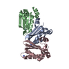
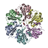
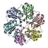
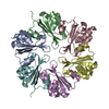
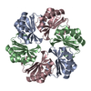
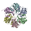
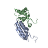
| Title | Structure of CcmK4 from Synechocystis sp. PCC6803 | ||||||
 Components Components | Carbon dioxide-concentrating mechanism protein CcmK homolog 4 | ||||||
 Keywords Keywords | STRUCTURAL PROTEIN / bacterial micro-compartment / carboxysome / CcmK4 / 2D assembly | ||||||
| Function / homology |  Function and homology information Function and homology informationstructural constituent of carboxysome shell / carboxysome / carbon fixation / photosynthesis Similarity search - Function | ||||||
| Biological species |  | ||||||
| Method |  X-RAY DIFFRACTION / SYNCHROTRON / MOLECULAR REPLACEMENT / Resolution: 1.8 Å X-RAY DIFFRACTION / SYNCHROTRON / MOLECULAR REPLACEMENT / Resolution: 1.8 Å | ||||||
 Authors Authors | Maveyraud, L. / Garcia-Alles, L.F. / Mourey, L. | ||||||
 Citation Citation | Journal: Plos One / Year: 2019 Title: Occurrence and stability of hetero-hexamer associations formed by beta-carboxysome CcmK shell components. Authors: Garcia-Alles, L.F. / Root, K. / Maveyraud, L. / Aubry, N. / Lesniewska, E. / Mourey, L. / Zenobi, R. / Truan, G. | ||||||
| History |
|
- Structure visualization
| Structure viewer | Molecule: MolmilJmol/JSmol |
|---|
- Downloads & links
-Download
| PDBx/mmCIF format | 6scr.cif.gz | 131 KB | Display | PDBx/mmCIF format |
|---|---|---|---|---|
| PDB format | pdb6scr.ent.gz | 102.4 KB | Display | PDB format |
| PDBx/mmJSON format | 6scr.json.gz | Tree view | PDBx/mmJSON format | |
| Others |  Other downloads Other downloads |
-Validation report
| Arichive directory | https://data.pdbj.org/pub/pdb/validation_reports/sc/6scrftp://data.pdbj.org/pub/pdb/validation_reports/sc/6scr | HTTPS FTP |
|---|
-Related structure data
| Related structure data |  2a18S S: Starting model for refinement |
|---|---|
| Similar structure data |
-Links
 PDBj
PDBj

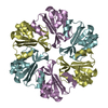
- Assembly
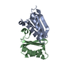
| Deposited unit | 
| ||||||||||||
|---|---|---|---|---|---|---|---|---|---|---|---|---|---|
| 1 | 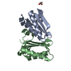
| ||||||||||||
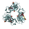
| Unit cell |
| ||||||||||||
| Components on special symmetry positions |
|
-Components
| #1: Protein | Mass: 13863.758 Da / Num. of mol.: 2 / Mutation: MAHHHHASGENLYFQGAMA is an added Nterminal tag Source method: isolated from a genetically manipulated source Source: (gene. exp.) Strain: PCC 6803 / Kazusa / Gene: ccmK4, slr1839 / Plasmid: pET-26b Production host: References: UniProt: P73407 #2: Chemical | ChemComp-EDO / |   Mass: 62.068 Da / Num. of mol.: 1 / Source method: obtained synthetically / Formula: C2H6O2 Mass: 62.068 Da / Num. of mol.: 1 / Source method: obtained synthetically / Formula: C2H6O2#3: Water | ChemComp-HOH / |  Mass: 18.015 Da / Num. of mol.: 147 / Source method: isolated from a natural source / Formula: H2O Mass: 18.015 Da / Num. of mol.: 147 / Source method: isolated from a natural source / Formula: H2OHas ligand of interest | N | |
|---|
-Experimental details
-Experiment
| Experiment | Method: X-RAY DIFFRACTION / Number of used crystals: 1 |
|---|
- Sample preparation
| Crystal | Density Matthews: 2.13 Å3/Da / Density % sol: 42.27 % |
|---|---|
| Crystal grow | Temperature: 285 K / Method: vapor diffusion, sitting drop / pH: 4.6 Details: 22 % (w/v) PEG 4000 0.2 M ammonium sulfate 0.1 M sodium acetate at pH 4.6 |
-Data collection
| Diffraction | Mean temperature: 100 K / Serial crystal experiment: N | ||||||||||||||||||||||||||||||
|---|---|---|---|---|---|---|---|---|---|---|---|---|---|---|---|---|---|---|---|---|---|---|---|---|---|---|---|---|---|---|---|
| Diffraction source | Source: SYNCHROTRON / Site: ESRF  / Beamline: MASSIF-3 / Wavelength: 0.9677 Å / Beamline: MASSIF-3 / Wavelength: 0.9677 Å | ||||||||||||||||||||||||||||||
| Detector | Type: DECTRIS EIGER X 4M / Detector: PIXEL / Date: Jun 28, 2018 | ||||||||||||||||||||||||||||||
| Radiation | Protocol: SINGLE WAVELENGTH / Monochromatic (M) / Laue (L): M / Scattering type: x-ray | ||||||||||||||||||||||||||||||
| Radiation wavelength | Wavelength: 0.9677 Å / Relative weight: 1 | ||||||||||||||||||||||||||||||
| Reflection | Resolution: 1.8→61.21 Å / Num. obs: 17065 / % possible obs: 99.6 % / Redundancy: 10.5 % / CC1/2: 0.999 / Rmerge(I) obs: 0.095 / Rpim(I) all: 0.031 / Rrim(I) all: 0.1 / Χ2: 0.97 / Net I/σ(I): 14.1 | ||||||||||||||||||||||||||||||
| Reflection shell | Diffraction-ID: 1
|
- Processing
| Software |
| ||||||||||||||||||||
|---|---|---|---|---|---|---|---|---|---|---|---|---|---|---|---|---|---|---|---|---|---|
| Refinement | Method to determine structure: MOLECULAR REPLACEMENT Starting model: 2A18 Resolution: 1.8→61.2 Å / Cross valid method: FREE R-VALUE / Phase error: 27.08
| ||||||||||||||||||||
| Solvent computation | Shrinkage radii: 0.9 Å / VDW probe radii: 1.11 Å | ||||||||||||||||||||
| Displacement parameters | Biso mean: 32.7 Å2 | ||||||||||||||||||||
| Refinement step | Cycle: LAST / Resolution: 1.8→61.2 Å
|
