 Movie
Movie Controller


+ Open data

- Basic information
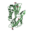
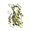
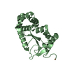
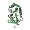
| Entry | Database: PDB / ID: 1pp0 | ||||||
|---|---|---|---|---|---|---|---|
| Title | volvatoxin A2 in monoclinic crystal | ||||||
 Components Components | volvatoxin A2 | ||||||
 Keywords Keywords | TOXIN / volvatoxin A2 / ingot crystal form | ||||||
| Function / homology |  Function and homology information Function and homology informationsporulation resulting in formation of a cellular spore / extracellular region Similarity search - Function | ||||||
| Biological species |  Volvariella volvacea (paddy straw mushroom) Volvariella volvacea (paddy straw mushroom) | ||||||
| Method |  X-RAY DIFFRACTION / SYNCHROTRON / MOLECULAR REPLACEMENT / Resolution: 1.42 Å X-RAY DIFFRACTION / SYNCHROTRON / MOLECULAR REPLACEMENT / Resolution: 1.42 Å | ||||||
 Authors Authors | Lin, S.-C. / Lo, Y.-C. / Lin, J.-Y. / Liaw, Y.-C. | ||||||
 Citation Citation | Journal: J.Mol.Biol. / Year: 2004 Title: Crystal structures and electron micrographs of fungal volvatoxin A2 Authors: Lin, S.-C. / Lo, Y.-C. / Lin, J.-Y. / Liaw, Y.-C. | ||||||
| History |
|
- Structure visualization
| Structure viewer | Molecule: MolmilJmol/JSmol |
|---|
- Downloads & links
-Download
| PDBx/mmCIF format | 1pp0.cif.gz | 181.5 KB | Display | PDBx/mmCIF format |
|---|---|---|---|---|
| PDB format | pdb1pp0.ent.gz | 142.5 KB | Display | PDB format |
| PDBx/mmJSON format | 1pp0.json.gz | Tree view | PDBx/mmJSON format | |
| Others |  Other downloads Other downloads |
-Validation report
| Arichive directory | https://data.pdbj.org/pub/pdb/validation_reports/pp/1pp0ftp://data.pdbj.org/pub/pdb/validation_reports/pp/1pp0 | HTTPS FTP |
|---|
-Related structure data
| Related structure data | 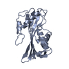 1pp6C  1vcyC 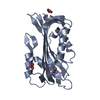 1vgfC  1iw3 C: citing same article ( S: Starting model for refinement |
|---|---|
| Similar structure data |


-Links
 PDBj
PDBj

- Assembly
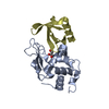
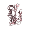
| Deposited unit | 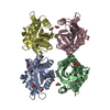
| ||||||||
|---|---|---|---|---|---|---|---|---|---|
| 1 | 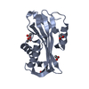
| ||||||||
| 2 | 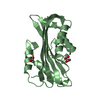
| ||||||||
| 3 | 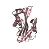
| ||||||||
| 4 | 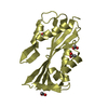
| ||||||||
| Unit cell |
|
-Components
| #1: Protein | Mass: 22356.961 Da / Num. of mol.: 4 / Source method: isolated from a natural source Source: (natural) Volvariella volvacea (paddy straw mushroom)References: UniProt: Q6USC4 #2: Chemical | ChemComp-ACY /   Mass: 60.052 Da / Num. of mol.: 13 / Source method: obtained synthetically / Formula: C2H4O2 Mass: 60.052 Da / Num. of mol.: 13 / Source method: obtained synthetically / Formula: C2H4O2#3: Water | ChemComp-HOH / |  Mass: 18.015 Da / Num. of mol.: 756 / Source method: isolated from a natural source / Formula: H2O Mass: 18.015 Da / Num. of mol.: 756 / Source method: isolated from a natural source / Formula: H2O |
|---|
-Experimental details
-Experiment
| Experiment | Method: X-RAY DIFFRACTION / Number of used crystals: 1 |
|---|
- Sample preparation
| Crystal | Density Matthews: 2.27 Å3/Da / Density % sol: 45.37 % |
|---|---|
| Crystal grow | Temperature: 298 K / Method: vapor diffusion, hanging drop Details: ammonium sulfate, PEG 4000, acetate buffer, VAPOR DIFFUSION, HANGING DROP, temperature 298K |
-Data collection
| Diffraction | Mean temperature: 133 K |
|---|---|
| Diffraction source | Source: SYNCHROTRON / Site: SPring-8  / Beamline: BL38B1 / Wavelength: 0.9236 Å / Beamline: BL38B1 / Wavelength: 0.9236 Å |
| Detector | Type: ADSC QUANTUM 4 / Detector: CCD / Date: Nov 1, 2001 |
| Radiation | Protocol: SINGLE WAVELENGTH / Monochromatic (M) / Laue (L): M / Scattering type: x-ray |
| Radiation wavelength | Wavelength: 0.9236 Å / Relative weight: 1 |
| Reflection | Resolution: 1.4→29.85 Å / Num. all: 172073 / Num. obs: 146871 / % possible obs: 85.4 % / Observed criterion σ(I): -3 / Redundancy: 5.8 % / Biso Wilson estimate: 24.5 Å2 / Limit h max: 81 / Limit h min: -108 / Limit k max: 41 / Limit k min: -108 / Limit l max: 81 / Limit l min: 0 / Observed criterion F max: 895251.86 / Observed criterion F min: 0.32 / Rmerge(I) obs: 0.036 / Net I/σ(I): 26 |
| Reflection shell | Resolution: 1.4→1.42 Å / Rmerge(I) obs: 0.258 / Mean I/σ(I) obs: 2 / % possible all: 49.1 |
- Processing
| Software |
| ||||||||||||||||||||||||||||||||||||||||||||||||||||||||||||||||||||||||||||||||||||||||||
|---|---|---|---|---|---|---|---|---|---|---|---|---|---|---|---|---|---|---|---|---|---|---|---|---|---|---|---|---|---|---|---|---|---|---|---|---|---|---|---|---|---|---|---|---|---|---|---|---|---|---|---|---|---|---|---|---|---|---|---|---|---|---|---|---|---|---|---|---|---|---|---|---|---|---|---|---|---|---|---|---|---|---|---|---|---|---|---|---|---|---|---|
| Refinement | Method to determine structure: MOLECULAR REPLACEMENT Starting model: 1IW3 1iw3 Resolution: 1.42→29.85 Å / Rfactor Rfree error: 0.003 / Occupancy max: 1 / Occupancy min: 1 / Isotropic thermal model: anisotropic / Cross valid method: THROUGHOUT / σ(F): 0 / σ(I): 0 / Stereochemistry target values: Engh & Huber
| ||||||||||||||||||||||||||||||||||||||||||||||||||||||||||||||||||||||||||||||||||||||||||
| Solvent computation | Solvent model: CNS bulk solvent model used / Bsol: 68.7373 Å2 / ksol: 0.394891 e/Å3 | ||||||||||||||||||||||||||||||||||||||||||||||||||||||||||||||||||||||||||||||||||||||||||
| Displacement parameters | Biso max: 116.37 Å2 / Biso mean: 30.32 Å2 / Biso min: 10.57 Å2
| ||||||||||||||||||||||||||||||||||||||||||||||||||||||||||||||||||||||||||||||||||||||||||
| Refine analyze |
| ||||||||||||||||||||||||||||||||||||||||||||||||||||||||||||||||||||||||||||||||||||||||||
| Refinement step | Cycle: LAST / Resolution: 1.42→29.85 Å
| ||||||||||||||||||||||||||||||||||||||||||||||||||||||||||||||||||||||||||||||||||||||||||
| Refine LS restraints |
| ||||||||||||||||||||||||||||||||||||||||||||||||||||||||||||||||||||||||||||||||||||||||||
| LS refinement shell | Refine-ID: X-RAY DIFFRACTION / Total num. of bins used: 8
| ||||||||||||||||||||||||||||||||||||||||||||||||||||||||||||||||||||||||||||||||||||||||||
| Xplor file |
|
