 Movie
Movie Controller

[English] 日本語
 Yorodumi
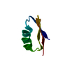
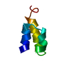
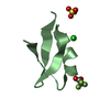
- PDB-4znf: HIGH-RESOLUTION THREE-DIMENSIONAL STRUCTURE OF A SINGLE ZINC FING... -
+ Open data

- Basic information
| Entry | Database: PDB / ID: 4znf | ||||||
|---|---|---|---|---|---|---|---|
| Title | HIGH-RESOLUTION THREE-DIMENSIONAL STRUCTURE OF A SINGLE ZINC FINGER FROM A HUMAN ENHANCER BINDING PROTEIN IN SOLUTION | ||||||
 Components Components | ZINC FINGER | ||||||
 Keywords Keywords | ZINC FINGER DNA BINDING DOMAIN | ||||||
| Function / homology |  Function and homology information Function and homology informationBMP signaling pathway / DNA-binding transcription repressor activity, RNA polymerase II-specific / sequence-specific double-stranded DNA binding / DNA-binding transcription factor activity, RNA polymerase II-specific / nuclear body / RNA polymerase II cis-regulatory region sequence-specific DNA binding / regulation of transcription by RNA polymerase II / negative regulation of transcription by RNA polymerase II / positive regulation of transcription by RNA polymerase II / mitochondrion ...BMP signaling pathway / DNA-binding transcription repressor activity, RNA polymerase II-specific / sequence-specific double-stranded DNA binding / DNA-binding transcription factor activity, RNA polymerase II-specific / nuclear body / RNA polymerase II cis-regulatory region sequence-specific DNA binding / regulation of transcription by RNA polymerase II / negative regulation of transcription by RNA polymerase II / positive regulation of transcription by RNA polymerase II / mitochondrion / zinc ion binding / nucleoplasm / nucleus / cytosol Similarity search - Function | ||||||
| Biological species |  Homo sapiens (human) Homo sapiens (human) | ||||||
| Method | SOLUTION NMR | ||||||
 Authors Authors | Gronenborn, A.M. / Clore, G.M. / Omichinski, J.G. | ||||||
 Citation Citation | Journal: Biochemistry / Year: 1990 Title: High-resolution three-dimensional structure of a single zinc finger from a human enhancer binding protein in solution. Authors: Omichinski, J.G. / Clore, G.M. / Appella, E. / Sakaguchi, K. / Gronenborn, A.M. | ||||||
| History |
|
- Structure visualization
| Structure viewer | Molecule: MolmilJmol/JSmol |
|---|
- Downloads & links
-Download
| PDBx/mmCIF format | 4znf.cif.gz | 447.3 KB | Display | PDBx/mmCIF format |
|---|---|---|---|---|
| PDB format | pdb4znf.ent.gz | 374.5 KB | Display | PDB format |
| PDBx/mmJSON format | 4znf.json.gz | Tree view | PDBx/mmJSON format | |
| Others |  Other downloads Other downloads |
-Validation report
| Arichive directory | https://data.pdbj.org/pub/pdb/validation_reports/zn/4znfftp://data.pdbj.org/pub/pdb/validation_reports/zn/4znf | HTTPS FTP |
|---|
-Related structure data


-Links
 PDBj
PDBj


- Assembly
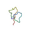
| Deposited unit | 
| |||||||||
|---|---|---|---|---|---|---|---|---|---|---|
| 1 |
| |||||||||
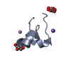
| Atom site foot note | 1: THE C-TERMINAL TWO RESIDUES, LYS 29 AND LYS 30, ARE ILL-DEFINED. | |||||||||
| NMR ensembles |
|
-Components
| #1: Protein/peptide | Mass: 3569.212 Da / Num. of mol.: 1 Source method: isolated from a genetically manipulated source Source: (gene. exp.) Homo sapiens (human) / References: UniProt: P15822 |
|---|---|
| #2: Chemical | ChemComp-ZN /   Mass: 65.409 Da / Num. of mol.: 1 / Source method: obtained synthetically / Formula: Zn Mass: 65.409 Da / Num. of mol.: 1 / Source method: obtained synthetically / Formula: Zn |
-Experimental details
-Experiment
| Experiment | Method: SOLUTION NMR |
|---|
- Processing
| Refinement | Software ordinal: 1 Details: THE 3D STRUCTURE OF THE ZINC FINGER IN SOLUTION BY NMR IS BASED ON 487 APPROXIMATE INTERPROTON DISTANCE RESTRAINTS AND 63 TORSION ANGLE RESTRAINTS DERIVED FROM NOE AND COUPLING CONSTANT ...Details: THE 3D STRUCTURE OF THE ZINC FINGER IN SOLUTION BY NMR IS BASED ON 487 APPROXIMATE INTERPROTON DISTANCE RESTRAINTS AND 63 TORSION ANGLE RESTRAINTS DERIVED FROM NOE AND COUPLING CONSTANT MEASUREMENTS. THE STRUCTURES ARE CALCULATED USING THE HYBRID METRIC MATRIX DISTANCE GEOMETRY-DYNAMICAL SIMULATED ANNEALING METHOD DESCRIBED BY M. NILGES, G. M. CLORE, AND A. M. GRONENBORN (1988) FEBS LETT 229, 317. THIS ENTRY REPRESENTS 41 MODELS OF THE ZINC FINGER OF HUMAN ENHANCER BINDING PROTEIN. THE RESTRAINED MINIMIZED AVERAGE STRUCTURE (SA)$R DERIVED BY RESTRAINED LEAST SQUARE REFINEMENT OF THE MEAN STRUCTURE OBTAINED BY AVERAGING THE COORDINATES OF THE FINAL 41 SA STRUCTURES BEST FITTED TO EACH OTHER CAN BE FOUND IN PDB ENTRY 3ZNF. |
|---|---|
| NMR ensemble | Conformers submitted total number: 41 |
