 Movie
Movie Controller

[English] 日本語
 Yorodumi
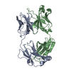
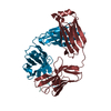
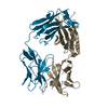
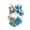
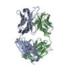
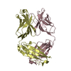
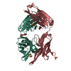
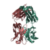
- PDB-4yo0: Crystal structure of monoclonal anti-human podoplanin antibody NZ... -
+ Open data

- Basic information
| Entry | Database: PDB / ID: 4yo0 | |||||||||
|---|---|---|---|---|---|---|---|---|---|---|
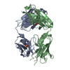
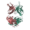
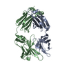
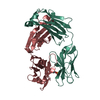
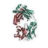
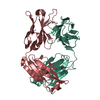
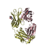
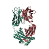
| Title | Crystal structure of monoclonal anti-human podoplanin antibody NZ-1 with bound PA peptide | |||||||||
 Components Components |
| |||||||||
 Keywords Keywords | IMMUNE SYSTEM / Antibodies / Monoclonal / Antibody Affinity / Chromatography / Affinity / Epitopes / Rats / Immunoglobulin Fab Fragments / Kinetics / Peptides / Protein Binding / Proteins / Recombinant Fusion Proteins / Human podoplanin | |||||||||
| Function / homology |  Function and homology information Function and homology informationregulation of myofibroblast contraction / lymphatic endothelial cell fate commitment / actin-mediated cell contraction / leading edge of lamellipodium / regulation of substrate adhesion-dependent cell spreading / chemokine binding / Specification of primordial germ cells / positive regulation of extracellular matrix disassembly / lymphangiogenesis / regulation of lamellipodium morphogenesis ...regulation of myofibroblast contraction / lymphatic endothelial cell fate commitment / actin-mediated cell contraction / leading edge of lamellipodium / regulation of substrate adhesion-dependent cell spreading / chemokine binding / Specification of primordial germ cells / positive regulation of extracellular matrix disassembly / lymphangiogenesis / regulation of lamellipodium morphogenesis / tetraspanin-enriched microdomain / positive regulation of platelet aggregation / filopodium membrane / wound healing, spreading of cells / anchoring junction / microvillus membrane / lamellipodium membrane / lymph node development / positive regulation of epithelial to mesenchymal transition / Rho protein signal transduction / GPVI-mediated activation cascade / ruffle / lung development / cell projection / filopodium / platelet activation / ruffle membrane / cell junction / cell migration / lamellipodium / regulation of cell shape / protein-folding chaperone binding / cytoplasmic vesicle / basolateral plasma membrane / cell adhesion / positive regulation of cell migration / apical plasma membrane / membrane raft / signaling receptor binding / negative regulation of cell population proliferation / negative regulation of apoptotic process / signal transduction / mitochondrion / membrane / plasma membrane / cytosol Similarity search - Function | |||||||||
| Biological species |   Homo sapiens (human) Homo sapiens (human) | |||||||||
| Method |  X-RAY DIFFRACTION / SYNCHROTRON / MOLECULAR REPLACEMENT / Resolution: 1.56 Å X-RAY DIFFRACTION / SYNCHROTRON / MOLECULAR REPLACEMENT / Resolution: 1.56 Å | |||||||||
 Authors Authors | Fujii, Y. / Kitago, Y. / Arimori, T. / Takagi, J. | |||||||||
 Citation Citation | Journal: J.Cell.Sci. / Year: 2016 Title: Tailored placement of a turn-forming PA tag into the structured domain of a protein to probe its conformational state Authors: Fujii, Y. / Matsunaga, Y. / Arimori, T. / Kitago, Y. / Ogasawara, S. / Kaneko, M.K. / Kato, Y. / Takagi, J. | |||||||||
| History |
|
- Structure visualization
| Structure viewer | Molecule: MolmilJmol/JSmol |
|---|
- Downloads & links
-Download
| PDBx/mmCIF format | 4yo0.cif.gz | 354.1 KB | Display | PDBx/mmCIF format |
|---|---|---|---|---|
| PDB format | pdb4yo0.ent.gz | 289.2 KB | Display | PDB format |
| PDBx/mmJSON format | 4yo0.json.gz | Tree view | PDBx/mmJSON format | |
| Others |  Other downloads Other downloads |
-Validation report
| Summary document | 4yo0_validation.pdf.gz | 488.3 KB | Display | wwPDB validaton report |
|---|---|---|---|---|
| Full document | 4yo0_full_validation.pdf.gz | 502.2 KB | Display | |
| Data in XML | 4yo0_validation.xml.gz | 38 KB | Display | |
| Data in CIF | 4yo0_validation.cif.gz | 55 KB | Display | |
| Arichive directory | https://data.pdbj.org/pub/pdb/validation_reports/yo/4yo0ftp://data.pdbj.org/pub/pdb/validation_reports/yo/4yo0 | HTTPS FTP |
-Related structure data
| Related structure data | 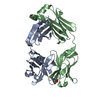 4ynySC S: Starting model for refinement C: citing same article ( |
|---|---|
| Similar structure data |


-Links
 PDBj
PDBj


- Assembly
| Deposited unit | 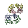
| ||||||||
|---|---|---|---|---|---|---|---|---|---|
| 1 | 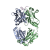
| ||||||||
| 2 | 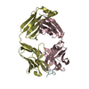
| ||||||||
| Unit cell |
|
-Components
| #1: Antibody | Mass: 25560.020 Da / Num. of mol.: 2 Source method: isolated from a genetically manipulated source Source: (gene. exp.) #2: Antibody | Mass: 25494.350 Da / Num. of mol.: 2 Source method: isolated from a genetically manipulated source Source: (gene. exp.) #3: Protein/peptide | Mass: 1345.432 Da / Num. of mol.: 2 / Source method: obtained synthetically / Source: (synth.) Homo sapiens (human) / References: UniProt: Q86YL7*PLUS#4: Chemical | ChemComp-EDO / |   Mass: 62.068 Da / Num. of mol.: 1 / Source method: obtained synthetically / Formula: C2H6O2 Mass: 62.068 Da / Num. of mol.: 1 / Source method: obtained synthetically / Formula: C2H6O2#5: Water | ChemComp-HOH / |  Mass: 18.015 Da / Num. of mol.: 499 / Source method: isolated from a natural source / Formula: H2O Mass: 18.015 Da / Num. of mol.: 499 / Source method: isolated from a natural source / Formula: H2OHas protein modification | Y | Sequence details | THE SEQUENCES OF THIS PROTEIN WERE NOT AVAILABLE AT THE UNIPROT KNOWLEDGEBASE DATABASE (UNIPROTKB) ...THE SEQUENCES OF THIS PROTEIN WERE NOT AVAILABLE AT THE UNIPROT KNOWLEDGEB | |
|---|
-Experimental details
-Experiment
| Experiment | Method: X-RAY DIFFRACTION / Number of used crystals: 1 |
|---|
- Sample preparation
| Crystal | Density Matthews: 2.11 Å3/Da / Density % sol: 41.59 % |
|---|---|
| Crystal grow | Temperature: 293 K / Method: vapor diffusion, hanging drop / Details: PEG 4000, sodium citrate |
-Data collection
| Diffraction | Mean temperature: 100 K |
|---|---|
| Diffraction source | Source: SYNCHROTRON / Site: Photon Factory  / Beamline: BL-17A / Wavelength: 0.98 Å / Beamline: BL-17A / Wavelength: 0.98 Å |
| Detector | Type: ADSC QUANTUM 270 / Detector: CCD / Date: Apr 20, 2013 |
| Radiation | Protocol: SINGLE WAVELENGTH / Monochromatic (M) / Laue (L): M / Scattering type: x-ray |
| Radiation wavelength | Wavelength: 0.98 Å / Relative weight: 1 |
| Reflection | Resolution: 1.56→40 Å / Num. obs: 121358 / % possible obs: 97.2 % / Redundancy: 7.5 % / Rsym value: 0.057 / Net I/σ(I): 17.5 |
| Reflection shell | Resolution: 1.56→1.65 Å / Redundancy: 7.2 % / Mean I/σ(I) obs: 1.5 / % possible all: 90.2 |
- Processing
| Software |
| |||||||||||||||||||||||||||||||||||||||||||||||||||||||||||||||||||||||||||||||||||||||||||||||||||||||||||||||||||||||||||||||||||||||||||||||||||||||||||||||||||||||||||||||||||||||||||||||||||||||||||||||||||||||||||||||||||||||||||||||||||||||||||||||||||||||||||||||||||
|---|---|---|---|---|---|---|---|---|---|---|---|---|---|---|---|---|---|---|---|---|---|---|---|---|---|---|---|---|---|---|---|---|---|---|---|---|---|---|---|---|---|---|---|---|---|---|---|---|---|---|---|---|---|---|---|---|---|---|---|---|---|---|---|---|---|---|---|---|---|---|---|---|---|---|---|---|---|---|---|---|---|---|---|---|---|---|---|---|---|---|---|---|---|---|---|---|---|---|---|---|---|---|---|---|---|---|---|---|---|---|---|---|---|---|---|---|---|---|---|---|---|---|---|---|---|---|---|---|---|---|---|---|---|---|---|---|---|---|---|---|---|---|---|---|---|---|---|---|---|---|---|---|---|---|---|---|---|---|---|---|---|---|---|---|---|---|---|---|---|---|---|---|---|---|---|---|---|---|---|---|---|---|---|---|---|---|---|---|---|---|---|---|---|---|---|---|---|---|---|---|---|---|---|---|---|---|---|---|---|---|---|---|---|---|---|---|---|---|---|---|---|---|---|---|---|---|---|---|---|---|---|---|---|---|---|---|---|---|---|---|---|---|---|---|---|---|---|---|---|---|---|---|---|---|---|---|---|---|---|---|---|---|---|---|---|---|---|---|---|---|---|---|---|---|---|---|
| Refinement | Method to determine structure: MOLECULAR REPLACEMENT Starting model: 4YNY Resolution: 1.56→40 Å / SU ML: 0.19 / Cross valid method: THROUGHOUT / σ(F): 1.34 / Phase error: 25.67
| |||||||||||||||||||||||||||||||||||||||||||||||||||||||||||||||||||||||||||||||||||||||||||||||||||||||||||||||||||||||||||||||||||||||||||||||||||||||||||||||||||||||||||||||||||||||||||||||||||||||||||||||||||||||||||||||||||||||||||||||||||||||||||||||||||||||||||||||||||
| Solvent computation | Shrinkage radii: 0.9 Å / VDW probe radii: 1.11 Å | |||||||||||||||||||||||||||||||||||||||||||||||||||||||||||||||||||||||||||||||||||||||||||||||||||||||||||||||||||||||||||||||||||||||||||||||||||||||||||||||||||||||||||||||||||||||||||||||||||||||||||||||||||||||||||||||||||||||||||||||||||||||||||||||||||||||||||||||||||
| Refinement step | Cycle: LAST / Resolution: 1.56→40 Å
| |||||||||||||||||||||||||||||||||||||||||||||||||||||||||||||||||||||||||||||||||||||||||||||||||||||||||||||||||||||||||||||||||||||||||||||||||||||||||||||||||||||||||||||||||||||||||||||||||||||||||||||||||||||||||||||||||||||||||||||||||||||||||||||||||||||||||||||||||||
| Refine LS restraints |
| |||||||||||||||||||||||||||||||||||||||||||||||||||||||||||||||||||||||||||||||||||||||||||||||||||||||||||||||||||||||||||||||||||||||||||||||||||||||||||||||||||||||||||||||||||||||||||||||||||||||||||||||||||||||||||||||||||||||||||||||||||||||||||||||||||||||||||||||||||
| LS refinement shell |
| |||||||||||||||||||||||||||||||||||||||||||||||||||||||||||||||||||||||||||||||||||||||||||||||||||||||||||||||||||||||||||||||||||||||||||||||||||||||||||||||||||||||||||||||||||||||||||||||||||||||||||||||||||||||||||||||||||||||||||||||||||||||||||||||||||||||||||||||||||
| Refinement TLS params. | Method: refined / Refine-ID: X-RAY DIFFRACTION
| |||||||||||||||||||||||||||||||||||||||||||||||||||||||||||||||||||||||||||||||||||||||||||||||||||||||||||||||||||||||||||||||||||||||||||||||||||||||||||||||||||||||||||||||||||||||||||||||||||||||||||||||||||||||||||||||||||||||||||||||||||||||||||||||||||||||||||||||||||
| Refinement TLS group |
|
