 Movie
Movie Controller


+ Open data

- Basic information
| Entry | Database: PDB / ID: 5itb | ||||||
|---|---|---|---|---|---|---|---|
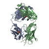
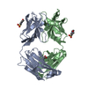
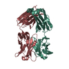
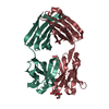
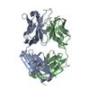
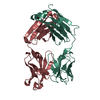
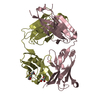
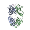
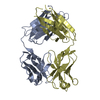
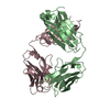
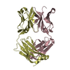
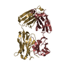
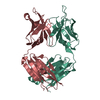
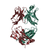
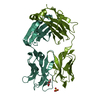
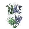
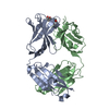
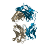
| Title | Crystal structure of the anti-RSV F Fab 14N4 | ||||||
 Components Components |
| ||||||
 Keywords Keywords | IMMUNE SYSTEM / antibody | ||||||
| Function / homology | Immunoglobulins / Immunoglobulin-like / Sandwich / Mainly Beta Function and homology information Function and homology information | ||||||
| Biological species |  Homo sapiens (human) Homo sapiens (human) | ||||||
| Method |  X-RAY DIFFRACTION / MOLECULAR REPLACEMENT / Resolution: 2 Å X-RAY DIFFRACTION / MOLECULAR REPLACEMENT / Resolution: 2 Å | ||||||
 Authors Authors | Mousa, J.J. / Crowe, J.E. | ||||||
 Citation Citation | Journal: Proc.Natl.Acad.Sci.USA / Year: 2016 Title: Structural basis for nonneutralizing antibody competition at antigenic site II of the respiratory syncytial virus fusion protein. Authors: Mousa, J.J. / Sauer, M.F. / Sevy, A.M. / Finn, J.A. / Bates, J.T. / Alvarado, G. / King, H.G. / Loerinc, L.B. / Fong, R.H. / Doranz, B.J. / Correia, B.E. / Kalyuzhniy, O. / Wen, X. / ...Authors: Mousa, J.J. / Sauer, M.F. / Sevy, A.M. / Finn, J.A. / Bates, J.T. / Alvarado, G. / King, H.G. / Loerinc, L.B. / Fong, R.H. / Doranz, B.J. / Correia, B.E. / Kalyuzhniy, O. / Wen, X. / Jardetzky, T.S. / Schief, W.R. / Ohi, M.D. / Meiler, J. / Crowe, J.E. | ||||||
| History |
|
- Structure visualization
| Structure viewer | Molecule: MolmilJmol/JSmol |
|---|
- Downloads & links
-Download
| PDBx/mmCIF format | 5itb.cif.gz | 105.7 KB | Display | PDBx/mmCIF format |
|---|---|---|---|---|
| PDB format | pdb5itb.ent.gz | 79.3 KB | Display | PDB format |
| PDBx/mmJSON format | 5itb.json.gz | Tree view | PDBx/mmJSON format | |
| Others |  Other downloads Other downloads |
-Validation report
| Arichive directory | https://data.pdbj.org/pub/pdb/validation_reports/it/5itbftp://data.pdbj.org/pub/pdb/validation_reports/it/5itb | HTTPS FTP |
|---|
-Related structure data
| Related structure data |  5j3dC  4q9qS S: Starting model for refinement C: citing same article ( |
|---|---|
| Similar structure data |


-Links
 PDBj
PDBj

- Assembly
| Deposited unit | 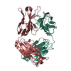
| ||||||||
|---|---|---|---|---|---|---|---|---|---|
| 1 |
| ||||||||
| Unit cell |
|
-Components
| #1: Antibody | Mass: 23128.938 Da / Num. of mol.: 1 Source method: isolated from a genetically manipulated source Source: (gene. exp.) Homo sapiens (human) / Production host: Homo sapiens (human) |
|---|---|
| #2: Antibody | Mass: 23764.240 Da / Num. of mol.: 1 Source method: isolated from a genetically manipulated source Source: (gene. exp.) Homo sapiens (human) / Production host: Homo sapiens (human) |
| #3: Water | ChemComp-HOH /  Mass: 18.015 Da / Num. of mol.: 416 / Source method: isolated from a natural source / Formula: H2O Mass: 18.015 Da / Num. of mol.: 416 / Source method: isolated from a natural source / Formula: H2O |
| Has protein modification | Y |
-Experimental details
-Experiment
| Experiment | Method: X-RAY DIFFRACTION / Number of used crystals: 1 |
|---|
- Sample preparation
| Crystal | Density Matthews: 2.18 Å3/Da / Density % sol: 43.58 % |
|---|---|
| Crystal grow | Temperature: 293 K / Method: vapor diffusion, sitting drop / Details: 20% PEG3350, 50 mM zinc acetate |
-Data collection
| Diffraction | Mean temperature: 80 K |
|---|---|
| Diffraction source | Source: ROTATING ANODE / Type: BRUKER AXS MICROSTAR / Wavelength: 1.54 Å |
| Detector | Type: Bruker Platinum 135 / Detector: CCD / Date: Sep 22, 2015 |
| Radiation | Protocol: SINGLE WAVELENGTH / Monochromatic (M) / Laue (L): M / Scattering type: x-ray |
| Radiation wavelength | Wavelength: 1.54 Å / Relative weight: 1 |
| Reflection | Resolution: 2→28.36 Å / Num. obs: 27310 / % possible obs: 99.6 % / Redundancy: 4.4 % / CC1/2: 0.991 / Rmerge(I) obs: 0.118 / Net I/σ(I): 8.8 |
| Reflection shell | Resolution: 2→2.07 Å / Rmerge(I) obs: 0.496 / Mean I/σ(I) obs: 2.2 / % possible all: 100 |
- Processing
| Software |
| |||||||||||||||||||||||||||||||||||||||||||||||||||||||||||||||||||||||||||||
|---|---|---|---|---|---|---|---|---|---|---|---|---|---|---|---|---|---|---|---|---|---|---|---|---|---|---|---|---|---|---|---|---|---|---|---|---|---|---|---|---|---|---|---|---|---|---|---|---|---|---|---|---|---|---|---|---|---|---|---|---|---|---|---|---|---|---|---|---|---|---|---|---|---|---|---|---|---|---|
| Refinement | Method to determine structure: MOLECULAR REPLACEMENT Starting model: 4Q9Q Resolution: 2→28.353 Å / SU ML: 0.19 / Cross valid method: FREE R-VALUE / σ(F): 1.35 / Phase error: 20.84 / Stereochemistry target values: ML
| |||||||||||||||||||||||||||||||||||||||||||||||||||||||||||||||||||||||||||||
| Solvent computation | Shrinkage radii: 0.9 Å / VDW probe radii: 1.11 Å / Solvent model: FLAT BULK SOLVENT MODEL | |||||||||||||||||||||||||||||||||||||||||||||||||||||||||||||||||||||||||||||
| Refinement step | Cycle: LAST / Resolution: 2→28.353 Å
| |||||||||||||||||||||||||||||||||||||||||||||||||||||||||||||||||||||||||||||
| Refine LS restraints |
| |||||||||||||||||||||||||||||||||||||||||||||||||||||||||||||||||||||||||||||
| LS refinement shell |
|
